9BT8 | pdb_00009bt8

Structure of Src in complex with beta-arrestin 1 revealing SH3 binding sites


Experimental Data Snapshot

  • Method: ELECTRON MICROSCOPY
  • Resolution: 3.34 Å
  • Aggregation State: PARTICLE 
  • Reconstruction Method: SINGLE PARTICLE 

Starting Models: experimental
View more details

wwPDB Validation   3D Report Full Report


This is version 1.2 of the entry. See complete history


Literature

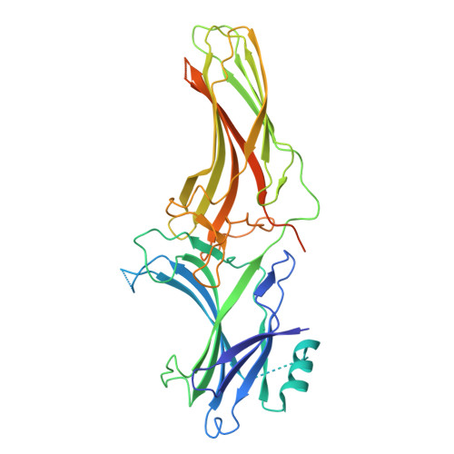
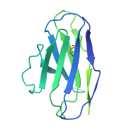
Mechanism of beta-arrestin 1 mediated Src activation via Src SH3 domain revealed by cryo-electron microscopy.

Pakharukova, N.Thomas, B.N.Bansia, H.Li, L.Bassford, D.K.Abzalimov, R.R.Kim, J.Kahsai, A.W.Pani, B.Xiao, K.Ochakovski, R.Liu, S.Zhang, X.Ahn, S.des Georges, A.Lefkowitz, R.J.

(2026) Nat Commun 

  • DOI: https://doi.org/10.1038/s41467-026-69884-1
  • Primary Citation of Related Structures:  
    9BT8, 9CX3, 9CX9

  • PubMed Abstract: 

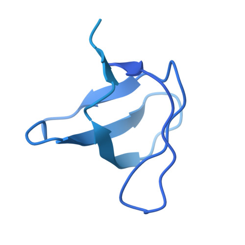
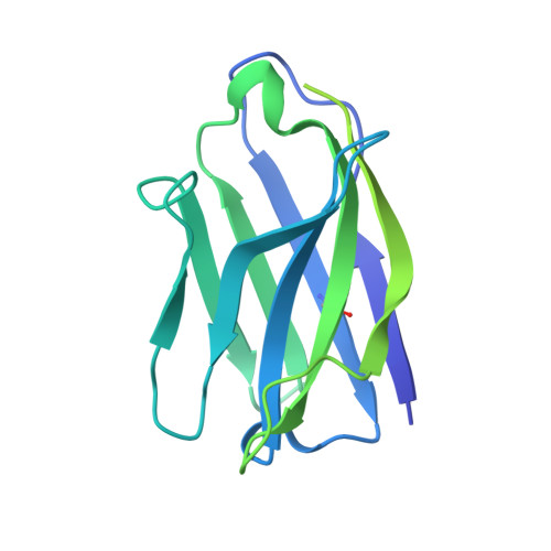
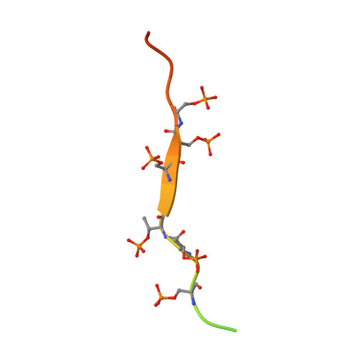
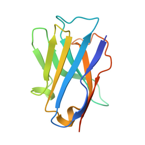
    Beta-arrestins (βarrs) are key regulators and transducers of G-protein coupled receptor signaling; however, little is known of how βarrs communicate with their downstream effectors. Here, we delineate structural mechanisms underlying βarr-mediated signal transduction. Using cryo-electron microscopy, we elucidate how βarr1 recruits and activates the non-receptor tyrosine kinase Src, a well-established signaling partner of βarrs. βarr1 engages Src SH3 through two distinct sites, each employing a different recognition mechanism: a polyproline motif in the N-domain and a non-proline-based interaction in the central crest region. At both sites βarr1 interacts with the aromatic surface of SH3, disrupting the autoinhibited conformation of Src and directly triggering its allosteric activation. This structural evidence establishes βarr1 as an active regulatory protein rather than a passive scaffold and suggests a potentially general mechanism for βarr-mediated signaling across diverse effectors.


  • Organizational Affiliation
    • Department of Medicine, Duke University Medical Center, Durham, NC, 27710, USA.

Macromolecules
Find similar proteins by:  (by identity cutoff)  |  3D Structure
Entity ID: 1
MoleculeChains Sequence LengthOrganismDetailsImage
Proto-oncogene tyrosine-protein kinase SrcA [auth C]477Gallus gallusMutation(s): 6 
Gene Names: SRC
EC: 2.7.10.2
UniProt
Find proteins for P00523 (Gallus gallus)
Explore P00523 
Go to UniProtKB:  P00523
Entity Groups  
Sequence Clusters30% Identity50% Identity70% Identity90% Identity95% Identity100% Identity
UniProt GroupP00523
Sequence Annotations
Expand
  • Reference Sequence
Find similar proteins by:  (by identity cutoff)  |  3D Structure
Entity ID: 2
MoleculeChains Sequence LengthOrganismDetailsImage
Antibody fragment Fab30, heavy chainB [auth H]237Bacteriophage sp.Mutation(s): 0 
Entity Groups  
Sequence Clusters30% Identity50% Identity70% Identity90% Identity95% Identity100% Identity
Sequence Annotations
Expand
  • Reference Sequence
Find similar proteins by:  (by identity cutoff)  |  3D Structure
Entity ID: 3
MoleculeChains Sequence LengthOrganismDetailsImage
Vasopressin V2 receptorC [auth V]29Homo sapiensMutation(s): 0 
UniProt & NIH Common Fund Data Resources
Find proteins for P30518 (Homo sapiens)
Explore P30518 
Go to UniProtKB:  P30518
PHAROS:  P30518
GTEx:  ENSG00000126895 
Entity Groups  
Sequence Clusters30% Identity50% Identity70% Identity90% Identity95% Identity100% Identity
UniProt GroupP30518
Sequence Annotations
Expand
  • Reference Sequence
Find similar proteins by:  (by identity cutoff)  |  3D Structure
Entity ID: 4
MoleculeChains Sequence LengthOrganismDetailsImage
Nanobody 32D [auth A]124Lama glamaMutation(s): 0 
Entity Groups  
Sequence Clusters30% Identity50% Identity70% Identity90% Identity95% Identity100% Identity
Sequence Annotations
Expand
  • Reference Sequence
Find similar proteins by:  (by identity cutoff)  |  3D Structure
Entity ID: 5
MoleculeChains Sequence LengthOrganismDetailsImage
Beta-arrestin-1E [auth B]392Rattus norvegicusMutation(s): 8 
Gene Names: Arrb1
UniProt
Find proteins for P29066 (Rattus norvegicus)
Explore P29066 
Go to UniProtKB:  P29066
Entity Groups  
Sequence Clusters30% Identity50% Identity70% Identity90% Identity95% Identity100% Identity
UniProt GroupP29066
Sequence Annotations
Expand
  • Reference Sequence
Find similar proteins by:  (by identity cutoff)  |  3D Structure
Entity ID: 6
MoleculeChains Sequence LengthOrganismDetailsImage
Antibody fragment Fab30, light chainF [auth L]215Bacteriophage sp.Mutation(s): 0 
Entity Groups  
Sequence Clusters30% Identity50% Identity70% Identity90% Identity95% Identity100% Identity
Sequence Annotations
Expand
  • Reference Sequence
Small Molecules
Modified Residues  2 Unique
IDChains TypeFormula2D DiagramParent
SEP
Query on SEP
C [auth V]L-PEPTIDE LINKINGC3 H8 N O6 PSER
TPO
Query on TPO
C [auth V]L-PEPTIDE LINKINGC4 H10 N O6 PTHR
Experimental Data & Validation

Experimental Data

  • Method: ELECTRON MICROSCOPY
  • Resolution: 3.34 Å
  • Aggregation State: PARTICLE 
  • Reconstruction Method: SINGLE PARTICLE 
EM Software:
TaskSoftware PackageVersion
RECONSTRUCTIONcryoSPARC4.4.1
MODEL REFINEMENTPHENIX1.20.1-4487-000

Structure Validation

View Full Validation Report


Entry History & Funding Information

Deposition Data


Funding OrganizationLocationGrant Number
Howard Hughes Medical Institute (HHMI)United States--
National Institutes of Health/National Heart, Lung, and Blood Institute (NIH/NHLBI)United StatesHL016037-50
National Institutes of Health/National Institute of General Medical Sciences (NIH/NIGMS)United StatesGM133598
European Molecular Biology Organization (EMBO)European UnionALTF 1071-2017
Human Frontier Science Program (HFSP)FranceLT000174/2018

Revision History  (Full details and data files)

  • Version 1.0: 2024-11-13
    Type: Initial release
  • Version 1.1: 2024-11-27
    Changes: Data collection
  • Version 1.2: 2026-03-04
    Changes: Data collection, Database references