PARP enzyme de novo synthesis of protein-free poly(ADP-ribose).
Langelier, M.F., Mirhasan, M., Gilbert, K., Sverzhinksy, A., Furtos, A., Pascal, J.M.(2024) Mol Cell 84: 4758-4773.e6
- PubMed: 39536748 Search on PubMedSearch on PubMed Central
- DOI: https://doi.org/10.1016/j.molcel.2024.10.024
- Primary Citation Related Structures:
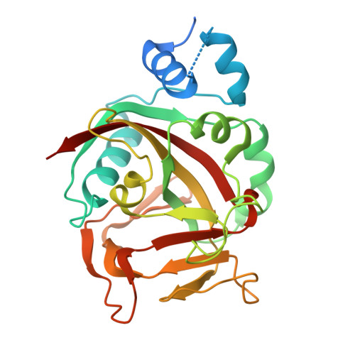
9BPY, 9DMC - PubMed Abstract:
PARP enzymes transfer ADP-ribose from NAD + onto proteins as a covalent modification that regulates multiple aspects of cell biology. Here, we identify an undiscovered catalytic activity for human PARP1: de novo generation of free PAR molecules that are not attached to proteins. Free PAR production arises when a molecule of NAD + or ADP-ribose docks in the PARP1 acceptor site and attaches to an NAD + molecule bound to the donor site, releasing nicotinamide and initiating ADP-ribose chains that emanate from NAD + /ADP-ribose rather than protein. Free PAR is also produced by human PARP2 and the PARP enzyme Tankyrase. We demonstrate that free PAR in cells is generated mostly by PARP1 de novo synthesis activity rather than by PAR-degrading enzymes PAR glycohydrolase (PARG), ARH3, and TARG1 releasing PAR from protein. The coincident production of free PAR and protein-linked modifications alters models for PAR signaling and broadens the scope of PARP enzyme signaling capacity.
- Department of Biochemistry and Molecular Medicine, Université de Montréal, Montréal, QC H3C 3J7, Canada.
Organizational Affiliation: