


Crystal Structure of UDP-N-acetylmuramoylalanine--D-glutamate ligase (MurD) from E. coli in complex with UMA and inhibitor A19
Liu, L., Seibold, S., Lovell, S., Battaile, K.P.To be published.
Experimental Data Snapshot
Starting Model: experimental
View more details
Entity ID: 1 | |||||
|---|---|---|---|---|---|
| Molecule | Chains | Sequence Length | Organism | Details | Image |
| UDP-N-acetylmuramoylalanine--D-glutamate ligase | 445 | Escherichia coli K-12 | Mutation(s): 0 Gene Names: murD EC: 6.3.2.9 | 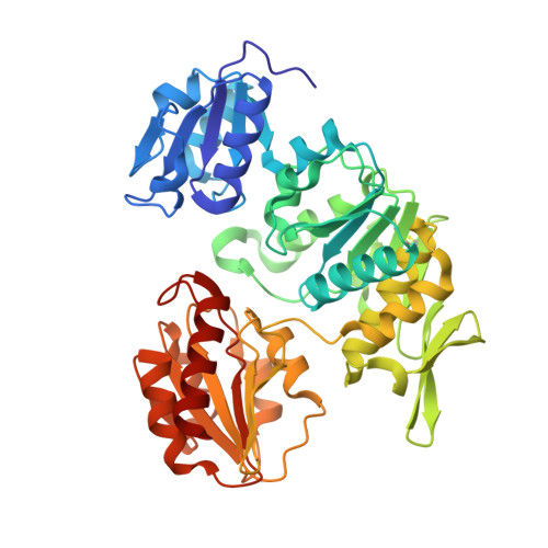 | |
UniProt | |||||
Entity Groups | |||||
| Sequence Clusters | 30% Identity50% Identity70% Identity90% Identity95% Identity100% Identity | ||||
| UniProt Group | P14900 | ||||
Sequence AnnotationsExpand | |||||
Reference Sequence | |||||
| Ligands 4 Unique | |||||
|---|---|---|---|---|---|
| ID | Chains | Name / Formula / InChI Key | 2D Diagram | 3D Interactions | |
| UMA Download:Ideal Coordinates CCD File | B [auth A] | URIDINE-5'-DIPHOSPHATE-N-ACETYLMURAMOYL-L-ALANINE C23 H36 N4 O20 P2 NTMMCWJNQNKACG-KBKUWGQMSA-N |  | ||
| A1AQS (Subject of Investigation/LOI) Download:Ideal Coordinates CCD File | C [auth A] | N-(4-{[(4S)-3-amino[1,2,4]triazolo[4,3-b]pyridazin-6-yl]sulfanyl}phenyl)acetamide C13 H12 N6 O S DDVAGHQEXKJINC-UHFFFAOYSA-N |  | ||
| SO4 Download:Ideal Coordinates CCD File | D [auth A] E [auth A] F [auth A] G [auth A] H [auth A] | SULFATE ION O4 S QAOWNCQODCNURD-UHFFFAOYSA-L |  | ||
| GOL Download:Ideal Coordinates CCD File | O [auth A] | GLYCEROL C3 H8 O3 PEDCQBHIVMGVHV-UHFFFAOYSA-N |  | ||
| Length ( Å ) | Angle ( ˚ ) |
|---|---|
| a = 65.488 | α = 90 |
| b = 65.488 | β = 90 |
| c = 134.764 | γ = 90 |
| Software Name | Purpose |
|---|---|
| PHENIX | refinement |
| Aimless | data scaling |
| XDS | data reduction |
| PHASER | phasing |
| Funding Organization | Location | Grant Number |
|---|---|---|
| National Institutes of Health/National Institute Of Allergy and Infectious Diseases (NIH/NIAID) | United States | 75N93022C00036 |
| National Institutes of Health/Office of the Director | United States | S10OD030394 |


