9BI0 | pdb_00009bi0

Streptomyces griseus Family 2B encapsulin shell with 2-methylisoborneol synthase cargo in 20 mM cAMP


Experimental Data Snapshot

  • Method: ELECTRON MICROSCOPY
  • Resolution: 2.93 Å
  • Aggregation State: PARTICLE 
  • Reconstruction Method: SINGLE PARTICLE 

Starting Model: in silico
View more details

wwPDB Validation 3D Report Full Report

Validation slider image for 9BI0

This is version 1.0 of the entry. See complete history

Literature

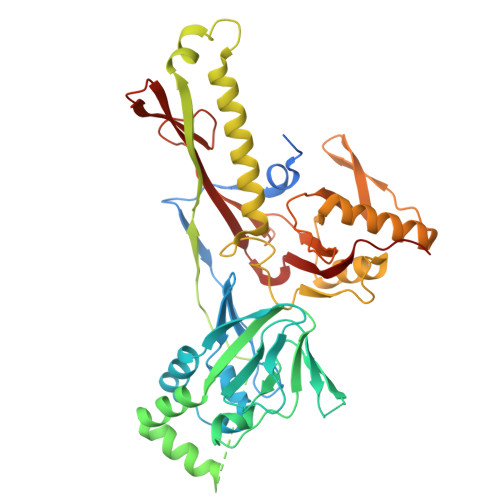
The biosynthesis of the odorant 2-methylisoborneol is compartmentalized inside a protein shell.

Andreas, M.P.Giessen, T.W.

(2024) Nat Commun 15: 9715-9715

  • DOI: https://doi.org/10.1038/s41467-024-54175-4
  • Primary Citation Related Structures: 
    9BHU, 9BHV, 9BI0

  • PubMed Abstract: 

    Terpenoids are the largest class of natural products, found across all domains of life. One of the most abundant bacterial terpenoids is the volatile odorant 2-methylisoborneol (2-MIB), partially responsible for the earthy smell of soil and musty taste of contaminated water. Many bacterial 2-MIB biosynthetic gene clusters were thought to encode a conserved transcription factor, named EshA in the model soil bacterium Streptomyces griseus. Here, we revise the function of EshA, now referred to as Sg Enc, and show that it is a Family 2B encapsulin shell protein. Using cryo-electron microscopy, we find that Sg Enc forms an icosahedral protein shell and encapsulates 2-methylisoborneol synthase (2-MIBS) as a cargo protein. Sg Enc contains a cyclic adenosine monophosphate (cAMP) binding domain (CBD)-fold insertion and a unique metal-binding domain, both displayed on the shell exterior. We show that Sg Enc CBDs do not bind cAMP. We find that 2-MIBS cargo loading is mediated by an N-terminal disordered cargo-loading domain and that 2-MIBS activity and Sg Enc shell structure are not modulated by cAMP. Our work redefines the function of EshA and establishes Family 2B encapsulins as cargo-loaded protein nanocompartments involved in secondary metabolite biosynthesis.


  • Organizational Affiliation
    • Department of Biological Chemistry, University of Michigan Medical School, Ann Arbor, MI, 48109, USA.

Macromolecule Content 

  • Total Structure Weight: 52.05 kDa 
  • Atom Count: 3,100 
  • Modeled Residue Count: 395 
  • Deposited Residue Count: 470 
  • Unique protein chains: 1

Macromolecules

Find similar proteins by:|  3D Structure
Entity ID: 1
MoleculeChains  Sequence LengthOrganismDetailsImage
Nucleotide-binding protein470Streptomyces griseus subsp. griseusMutation(s): 0 
Gene Names: mmpI_1NCTC13033_02226
UniProt
Find proteins for Q54255 (Streptomyces griseus)
Explore Q54255 
Go to UniProtKB:  Q54255
Entity Groups
Sequence Clusters30% Identity50% Identity70% Identity90% Identity95% Identity100% Identity
UniProt GroupQ54255
Sequence Annotations
Expand
Reference Sequence

Small Molecules

Ligands 1 Unique
IDChains Name / Formula / InChI Key2D Diagram3D Interactions
CA

Query on CA


Download:Ideal Coordinates CCD File
B [auth A]CALCIUM ION
Ca
BHPQYMZQTOCNFJ-UHFFFAOYSA-N

Experimental Data & Validation

Experimental Data

  • Method: ELECTRON MICROSCOPY
  • Resolution: 2.93 Å
  • Aggregation State: PARTICLE 
  • Reconstruction Method: SINGLE PARTICLE 
EM Software:
TaskSoftware PackageVersion
RECONSTRUCTIONcryoSPARC4.4.0+231114
MODEL REFINEMENTCoot8.9.1
MODEL REFINEMENTPHENIX1.20.1-4487

Structure Validation

View Full Validation Report


Entry History 

& Funding Information

Deposition Data


Funding OrganizationLocationGrant Number
National Institutes of Health/National Institute of General Medical Sciences (NIH/NIGMS)United StatesR35GM133325

Revision History  (Full details and data files)

  • Version 1.0: 2024-12-04
    Type: Initial release