9BDF | pdb_00009bdf

Influenza A virus Hemagglutinin H3/Darwin/6/2021 in complex with Fab ADI-85666


Experimental Data Snapshot

  • Method: ELECTRON MICROSCOPY
  • Resolution: 3.01 Å
  • Aggregation State: PARTICLE 
  • Reconstruction Method: SINGLE PARTICLE 

wwPDB Validation 3D Report Full Report

Validation slider image for 9BDF

This is version 1.1 of the entry. See complete history

Literature

Conserved sites on the influenza H1 and H3 hemagglutinin recognized by human antibodies.

Maurer, D.P.Vu, M.Ferreira Ramos, A.S.Dugan, H.L.Khalife, P.Geoghegan, J.C.Walker, L.M.Bajic, G.Schmidt, A.G.

(2025) Sci Adv 11: eadu9140-eadu9140

  • DOI: https://doi.org/10.1126/sciadv.adu9140
  • Primary Citation Related Structures: 
    9BDF, 9BDG

  • PubMed Abstract: 

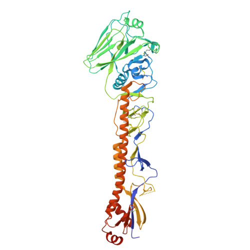
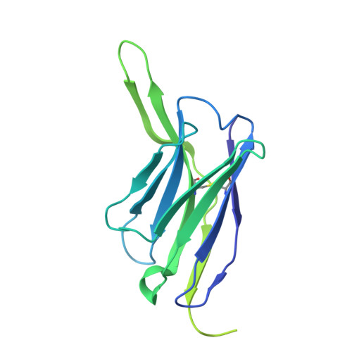
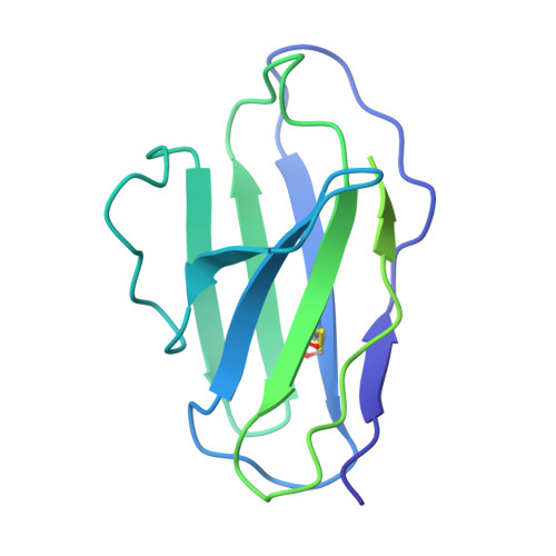
    Monoclonal antibodies (mAbs) targeting the influenza hemagglutinin (HA) can be used as prophylactics or templates for next-generation vaccines. Here, we isolated broad, subtype-neutralizing mAbs from human B cells recognizing the H1 or H3 HA "head" and a mAb engaging the conserved stem. The H1 mAbs bind the lateral patch epitope on HAs from 1933 to 2021 and a prepandemic swine H1N1 virus. We improved neutralization potency using directed evolution toward a contemporary H1 HA. Deep mutational scanning of four antigenically distinct H1N1 viruses identified potential viral escape pathways. For the H3 mAbs, we used cryo-electron microscopy to define their epitopes: One mAb binds the side of the HA head, accommodating the N133 glycan and a pocket underneath the receptor binding site; the other mAb recognizes an HA stem epitope that partially overlaps with previously characterized mAbs but with distinct antibody variable genes. Collectively, these mAbs identify conserved sites recognized by broadly-reactive mAbs that may be elicited by next-generation vaccines.


  • Organizational Affiliation
    • Ragon Institute of Mass General, MIT, and Harvard, Cambridge, MA 02139, USA.

Macromolecule Content 

  • Total Structure Weight: 327.9 kDa 
  • Atom Count: 17,167 
  • Modeled Residue Count: 2,126 
  • Deposited Residue Count: 2,904 
  • Unique protein chains: 3

Macromolecules

Find similar proteins by:|  3D Structure
Entity ID: 1
MoleculeChains  Sequence LengthOrganismDetailsImage
HemagglutininA,
F [auth G],
G [auth H]
514Influenza A virusMutation(s): 0 
Gene Names: HA
UniProt
Find proteins for A0A8F5JT24 (Influenza A virus)
Explore A0A8F5JT24 
Go to UniProtKB:  A0A8F5JT24
Entity Groups
Sequence Clusters30% Identity50% Identity70% Identity90% Identity95% Identity100% Identity
UniProt GroupA0A8F5JT24
Glycosylation
Glycosylation Sites: 8
Sequence Annotations
Expand
Reference Sequence
Find similar proteins by:|  3D Structure
Entity ID: 2
MoleculeChains  Sequence LengthOrganismDetailsImage
ADI-85666 Fab heavy chain
B, C, E
240Homo sapiensMutation(s): 0 
Find similar proteins by:|  3D Structure
Entity ID: 3
MoleculeChains  Sequence LengthOrganismDetailsImage
ADI-85666 Fab light chainD,
H [auth I],
I [auth J]
214Homo sapiensMutation(s): 0 

Oligosaccharides

Help  
Entity ID: 4
MoleculeChains Length2D Diagram GlycosylationD Interactions
alpha-D-mannopyranose-(1-3)-beta-D-mannopyranose-(1-4)-2-acetamido-2-deoxy-beta-D-glucopyranose-(1-4)-2-acetamido-2-deoxy-beta-D-glucopyranoseJ [auth F],
M,
R
4N-Glycosylation
Glycosylation Resources
GlyTouCan: G81315DD
GlyCosmos: G81315DD
GlyGen: G81315DD
Entity ID: 5
MoleculeChains Length2D Diagram GlycosylationD Interactions
2-acetamido-2-deoxy-beta-D-glucopyranose-(1-4)-2-acetamido-2-deoxy-beta-D-glucopyranose
K, N, O, P, Q
2N-Glycosylation
Glycosylation Resources
GlyTouCan: G42666HT
GlyCosmos: G42666HT
GlyGen: G42666HT
Entity ID: 6
MoleculeChains Length2D Diagram GlycosylationD Interactions
beta-D-mannopyranose-(1-4)-2-acetamido-2-deoxy-beta-D-glucopyranose-(1-4)-2-acetamido-2-deoxy-beta-D-glucopyranose
L
3N-Glycosylation
Glycosylation Resources
GlyTouCan: G15407YE
GlyCosmos: G15407YE
GlyGen: G15407YE

Small Molecules

Ligands 1 Unique
IDChains Name / Formula / InChI Key2D Diagram3D Interactions
NAG

Query on NAG


Download:Ideal Coordinates CCD File
AA [auth G]
BA [auth G]
CA [auth G]
DA [auth H]
EA [auth H]
AA [auth G],
BA [auth G],
CA [auth G],
DA [auth H],
EA [auth H],
FA [auth H],
GA [auth H],
HA [auth H],
S [auth A],
T [auth A],
U [auth A],
V [auth A],
W [auth A],
X [auth G],
Y [auth G],
Z [auth G]
2-acetamido-2-deoxy-beta-D-glucopyranose
C8 H15 N O6
OVRNDRQMDRJTHS-FMDGEEDCSA-N

Experimental Data & Validation

Experimental Data

  • Method: ELECTRON MICROSCOPY
  • Resolution: 3.01 Å
  • Aggregation State: PARTICLE 
  • Reconstruction Method: SINGLE PARTICLE 
EM Software:
TaskSoftware PackageVersion
MODEL REFINEMENTPHENIX1.21_5207

Structure Validation

View Full Validation Report


Entry History 

& Funding Information

Deposition Data


Funding OrganizationLocationGrant Number
National Institutes of Health/National Institute Of Allergy and Infectious Diseases (NIH/NIAID)United States75N93019C00051

Revision History  (Full details and data files)

  • Version 1.0: 2024-11-13
    Type: Initial release
  • Version 1.1: 2025-05-28
    Changes: Data collection, Database references