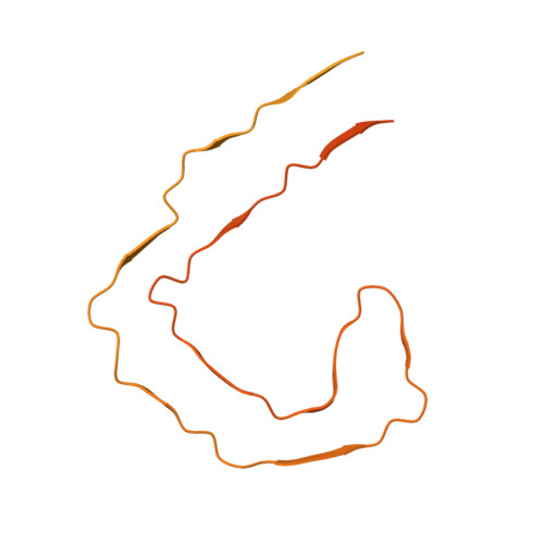
Milligram-scale assembly and NMR fingerprint of tau fibrils adopting the Alzheimer's disease fold.
Duan, P., El Mammeri, N., Hong, M.(2024) J Biological Chem 300: 107326-107326
- PubMed: 38679331
- DOI: https://doi.org/10.1016/j.jbc.2024.107326
- Primary Citation of Related Structures:
9BBL, 9BBM - PubMed Abstract:
In the Alzheimer's disease (AD) brain, the microtubule-associated protein tau aggregates into paired helical filaments (PHFs) in which each protofilament has a C-shaped conformation. In vitro assembly of tau fibrils adopting this fold is highly valuable for both fundamental and applied studies of AD without requiring patient-brain extracted fibrils. To date, reported methods for forming AD-fold tau fibrils have been irreproducible and sensitive to subtle variations in fibrillization conditions. Here we describe a route to reproducibly assemble tau fibrils adopting the AD fold on the multi-milligram scale. We investigated the fibrilization conditions of two constructs, and found that a tau (297-407) construct that contains four AD phospho-mimetic glutamate mutations robustly formed the C-shaped conformation. Two- and three-dimensional correlation solid-state NMR spectra show a single predominant set of chemical shifts, indicating a single molecular conformation. Negative-stain electron microscopy and cryoelectron microscopy data confirm that the protofilament formed by 4E-tau (297-407) adopts the C-shaped conformation, which associates into paired, triple and quadruple helical filaments. In comparison, NMR spectra indicate that a previously reported construct, tau (297-391), forms a mixture of a four-layered dimer structure and the C-shaped structure, whose populations are highly sensitive to the environmental conditions. The determination of the NMR chemical shifts of the AD-fold tau opens the possibility for future studies of tau fibril conformations and ligand binding by NMR. The quantitative assembly of tau fibrils adopting the AD fold should facilitate the development of diagnostic and therapeutic compounds that target AD tau.
Organizational Affiliation:
Department of Chemistry, Massachusetts Institute of Technology, 170 Albany Street, Cambridge, MA 02139.


