9BBF | pdb_00009bbf

Structure of Clostridioides difficile Component A (50-463) in Complex with a CDTb Oligomer


Experimental Data Snapshot

  • Method: ELECTRON MICROSCOPY
  • Resolution: 3.60 Å
  • Aggregation State: PARTICLE 
  • Reconstruction Method: SINGLE PARTICLE 

wwPDB Validation 3D Report Full Report

Validation slider image for 9BBF

This is version 1.2 of the entry. See complete history

Literature

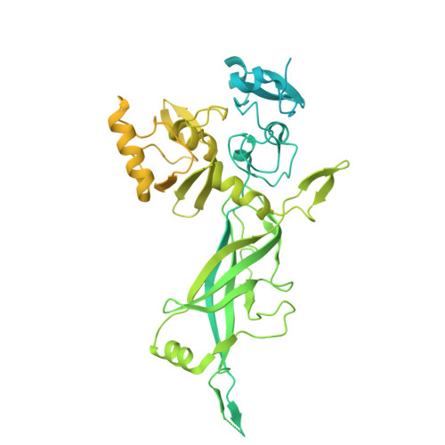
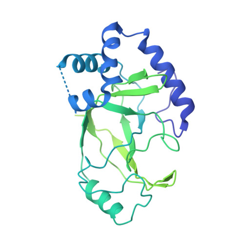
The N-terminus of the Clostridioides difficile transferase A component directs toxin activity and potency.

Mullard, R.M.Sheedlo, M.J.

(2025) mBio 16: e0240524-e0240524

  • DOI: https://doi.org/10.1128/mbio.02405-24
  • Primary Citation Related Structures: 
    9BBF

  • PubMed Abstract: 

    Clostridioides difficile infection is the leading cause of antibiotic-associated, hospital-acquired diarrhea in the USA; the pathology of which is mediated by toxins. The presence of a toxin known as the C. difficile Transferase (CDT) in some clinical isolates is linked to severe symptoms including increased incidence of reinfection and higher rates of mortality. Despite its apparent importance to C. difficile pathology, a mechanistic model of how CDT intoxicates cells remains incomplete. Here, we describe a motif composed of acidic and basic residues (the KDKEK motif) that is essential for toxin function. Using Cryogenic Electron Microscopy (Cryo-EM), we highlight an orientation of the KDKEK motif wherein the acidic residues engage structures thought to play an important role during toxin delivery. We thus present a model wherein these interactions prime CDT for entry into host cells. We expect that this model can be extrapolated to other bacterial toxins to understand how they enter cells.IMPORTANCE Clostridioides difficile is the leading cause of hospital-acquired infectious diarrhea in the USA. The pathology that accompanies infection is triggered by toxins produced by the bacterium. One of these, the C. difficile Transferase (CDT), has been associated with poorer patient outcomes, although a direct connection to CDT activity has remained elusive. Herein, we present new insight into the mechanism of CDT intoxication and define two regions of the toxin as important for its activity. Moreover, we have generated mutants of CDT that retain the ability to assemble but can no longer intoxicate host cells. In the future, we expect these mutants will serve as valuable tools to help elucidate the role of CDT during infection.


  • Organizational Affiliation
    • Department of Pharmacology, University of Minnesota Medical School, Minneapolis, Minnesota, USA.

Macromolecule Content 

  • Total Structure Weight: 708.21 kDa 
  • Atom Count: 19,880 
  • Modeled Residue Count: 2,533 
  • Deposited Residue Count: 6,259 
  • Unique protein chains: 2

Macromolecules

Find similar proteins by:|  3D Structure
Entity ID: 1
MoleculeChains  Sequence LengthOrganismDetailsImage
ADP-ribosyltransferase binding component
A, B, C, D, E
A, B, C, D, E, F, G
835Clostridioides difficileMutation(s): 0 
Gene Names: cdtB
UniProt
Find proteins for A8DS70 (Clostridioides difficile)
Explore A8DS70 
Go to UniProtKB:  A8DS70
Entity Groups
Sequence Clusters30% Identity50% Identity70% Identity90% Identity95% Identity100% Identity
UniProt GroupA8DS70
Sequence Annotations
Expand
Reference Sequence
Find similar proteins by:|  3D Structure
Entity ID: 2
MoleculeChains  Sequence LengthOrganismDetailsImage
ADP-ribosyltransferase enzymatic componentH [auth Z]414Clostridioides difficileMutation(s): 0 
Gene Names: cdtA
EC: 2.4.2
UniProt
Find proteins for Q9KH42 (Clostridioides difficile)
Explore Q9KH42 
Go to UniProtKB:  Q9KH42
Entity Groups
Sequence Clusters30% Identity50% Identity70% Identity90% Identity95% Identity100% Identity
UniProt GroupQ9KH42
Sequence Annotations
Expand
Reference Sequence

Small Molecules

Ligands 1 Unique
IDChains Name / Formula / InChI Key2D Diagram3D Interactions
CA

Query on CA


Download:Ideal Coordinates CCD File
I [auth A]
J [auth A]
K [auth B]
L [auth B]
M [auth C]
I [auth A],
J [auth A],
K [auth B],
L [auth B],
M [auth C],
N [auth C],
O [auth D],
P [auth D],
Q [auth E],
R [auth E],
S [auth F],
T [auth F],
U [auth G],
V [auth G]
CALCIUM ION
Ca
BHPQYMZQTOCNFJ-UHFFFAOYSA-N

Experimental Data & Validation

Experimental Data

  • Method: ELECTRON MICROSCOPY
  • Resolution: 3.60 Å
  • Aggregation State: PARTICLE 
  • Reconstruction Method: SINGLE PARTICLE 
EM Software:
TaskSoftware PackageVersion
MODEL REFINEMENTPHENIX

Structure Validation

View Full Validation Report


Entry History 

& Funding Information

Deposition Data


Funding OrganizationLocationGrant Number
National Institutes of Health/National Institute Of Allergy and Infectious Diseases (NIH/NIAID)United StatesR00AI154672

Revision History  (Full details and data files)

  • Version 1.0: 2024-11-27
    Type: Initial release
  • Version 1.1: 2025-05-21
    Changes: Data collection
  • Version 1.2: 2026-06-10
    Changes: Data collection, Database references