Interaction of CYP3A4 with the inhibitor cobicistat: Structural and mechanistic insights and comparison with ritonavir.
Sevrioukova, I.F.(2024) Arch Biochem Biophys 758: 110071-110071
- PubMed: 38909836 Search on PubMedSearch on PubMed Central
- DOI: https://doi.org/10.1016/j.abb.2024.110071
- Primary Citation Related Structures:
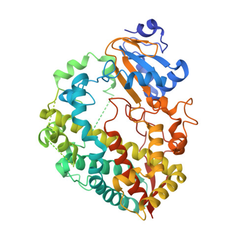
9BBB - PubMed Abstract:
Cobicistat is a derivative of ritonavir marketed as a pharmacoenhancer for anti-HIV therapy. This study investigated the interaction of cobicistat with the target protein, drug-metabolizing cytochrome P450 3A4 (CYP3A4), at the molecular level using spectral, kinetic, functional, and structural approaches. It was found that, similar to ritonavir, cobicistat directly coordinates to the heme via the thiazole nitrogen but its affinity and the binding rate are 2-fold lower: 0.030 μM and 0.72 s -1 , respectively. The newly determined 2.5 Å crystal structure of cobicistat-bound CYP3A4 suggests that these changes arise from the inability of cobicistat to H-bond to the active site S119 and establish multiple stabilizing contacts with the F-F' connecting fragment, which becomes disordered upon steric clashing with the bulky morpholine moiety. Nonetheless, cobicistat inhibits recombinant CYP3A4 as potently as ritonavir (IC 50 of 0.24 μM vs 0.22 μM, respectively) due to strong ligation to the heme and formation of extensive hydrophobic/aromatic interactions via the phenyl side-groups. To get insights into the inhibitory mechanism, the K257 residue, known to be solely and irreversibly modified by the reactive ritonavir metabolite, was substituted with alanine. Neither this nor control K266A mutation changed the extent of time-dependent inhibition of CYP3A4 by cobicistat and ritonavir, suggesting the existence of alternative inactivation mechanism(s). More importantly, K257 was found to be functionally important and contributed to CYP3A4 allosterism, possibly by modulating protein-ligand interactions through conformational dynamics.
- Department of Molecular Biology and Biochemistry, University of California, Irvine, CA, 92697-3900, USA. Electronic address: sevrioui@uci.edu.
Organizational Affiliation: