9BAJ | pdb_00009baj

Crystal structure of GDP-bound human K-RAS in a covalent complex with aryl sulfonyl fluoride compounds.


Experimental Data Snapshot

  • Method: X-RAY DIFFRACTION
  • Resolution: 1.49 Å
  • R-Value Free: 
    0.270 (Depositor), 0.270 (DCC) 
  • R-Value Work: 
    0.237 (Depositor), 0.239 (DCC) 
  • R-Value Observed: 
    0.239 (Depositor) 

Starting Model: experimental
View more details

wwPDB Validation   3D Report Full Report


Ligand Structure Quality Assessment 

Created with Raphaël 2.3.0Worse 01 BetterLigand structure goodness of fit to experimental dataBest fitted A1AK8Click on this verticalbar to view details

This is version 1.0 of the entry. See complete history


Literature

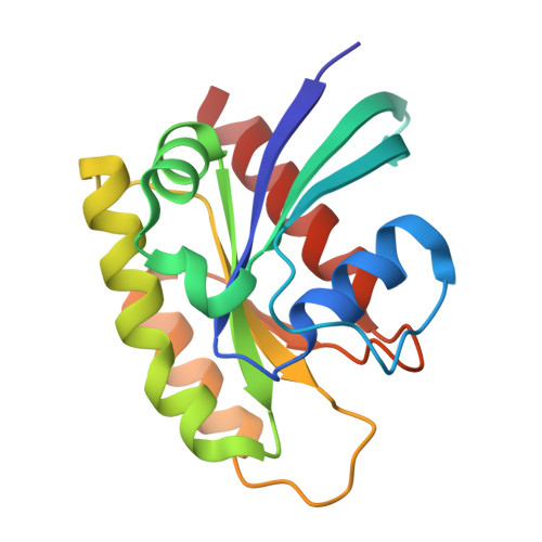
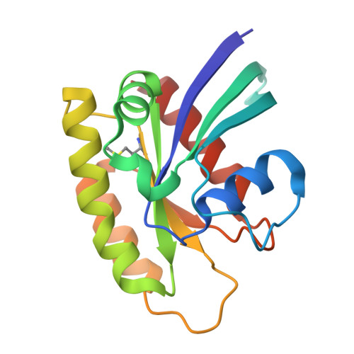
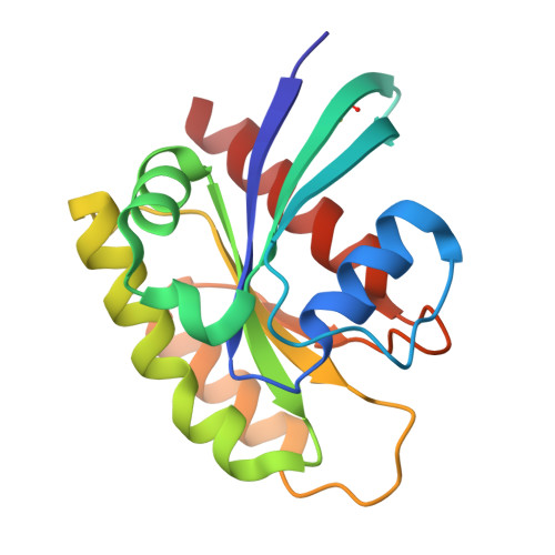
Small-Molecule KRAS Inhibitors by Tyrosine Covalent Bond Formation.

Landgraf, A.Brenner, R.Ghozayel, M.Bum-Erdene, K.Gonzalez-Gutierrez, G.Meroueh, S.

(2025) ChemMedChem : e202400624-e202400624

  • DOI: https://doi.org/10.1002/cmdc.202400624
  • Primary Citation of Related Structures:  
    9BAI, 9BAJ, 9BAK

  • PubMed Abstract: 

    The development of the KRAS G12C inhibitor sotorasib was a major advance towards drugging KRAS.  However, the G12C mutation is only found in about 10% of tumors with a KRAS mutation.  KRAS tyrosine amino acids could provide alternative sites for covalent drug development. Here, we screen a library of aryl sulfonyl fluorides to explore whether tyrosines on KRAS are accessible for covalent bond formation. We identify compound 1 (SOF-436), which inhibits KRAS nucleotide exchange by guanine exchange factor SOS1 and the binding of KRAS to effector protein RAF.  Tyr-64 was the major reaction site of 1 (SOF-436), although minor reaction at Tyr-71 was also observed. The fragment engages the Switch II pocket of KRAS based on mass spectrometry, nucleotide exchange, effector protein binding, nuclear magnetic resonance (NMR), and molecular dynamics simulations. Co-crystal structures of smaller fragments covalently bound to KRAS at Tyr-71 provide a strategy for the development of Switch I/II KRAS covalent inhibitors. A NanoBRET assay revealed that the compound and its analogs inhibit KRAS binding to RAF in mammalian cells. Although not yet suitable as chemical probes, these fragments provide starting points for small molecules to investigate tyrosine as a nucleophile for covalent inhibition of KRAS in tumors.


  • Organizational Affiliation

    Indiana University School of Medicine, Biochemistry, UNITED STATES OF AMERICA.


Macromolecules
Find similar proteins by:  (by identity cutoff)  |  3D Structure
Entity ID: 1
MoleculeChains Sequence LengthOrganismDetailsImage
Isoform 2B of GTPase KRas
A, D
169Homo sapiensMutation(s): 1 
Gene Names: KRASKRAS2RASK2
EC: 3.6.5.2
UniProt & NIH Common Fund Data Resources
Find proteins for P01116 (Homo sapiens)
Explore P01116 
Go to UniProtKB:  P01116
PHAROS:  P01116
GTEx:  ENSG00000133703 
Entity Groups  
Sequence Clusters30% Identity50% Identity70% Identity90% Identity95% Identity100% Identity
UniProt GroupP01116
Sequence Annotations
Expand
  • Reference Sequence
Find similar proteins by:  (by identity cutoff)  |  3D Structure
Entity ID: 2
MoleculeChains Sequence LengthOrganismDetailsImage
Isoform 2B of GTPase KRas169Homo sapiensMutation(s): 1 
Gene Names: KRASKRAS2RASK2
EC: 3.6.5.2
UniProt & NIH Common Fund Data Resources
Find proteins for P01116 (Homo sapiens)
Explore P01116 
Go to UniProtKB:  P01116
PHAROS:  P01116
GTEx:  ENSG00000133703 
Entity Groups  
Sequence Clusters30% Identity50% Identity70% Identity90% Identity95% Identity100% Identity
UniProt GroupP01116
Sequence Annotations
Expand
  • Reference Sequence
Find similar proteins by:  (by identity cutoff)  |  3D Structure
Entity ID: 3
MoleculeChains Sequence LengthOrganismDetailsImage
Isoform 2B of GTPase KRas169Homo sapiensMutation(s): 1 
Gene Names: KRASKRAS2RASK2
EC: 3.6.5.2
UniProt & NIH Common Fund Data Resources
Find proteins for P01116 (Homo sapiens)
Explore P01116 
Go to UniProtKB:  P01116
PHAROS:  P01116
GTEx:  ENSG00000133703 
Entity Groups  
Sequence Clusters30% Identity50% Identity70% Identity90% Identity95% Identity100% Identity
UniProt GroupP01116
Sequence Annotations
Expand
  • Reference Sequence
Small Molecules
Ligands 3 Unique
IDChains Name / Formula / InChI Key2D Diagram3D Interactions
GDP
Query on GDP

Download Ideal Coordinates CCD File 
G [auth A],
J [auth B],
M [auth C],
P [auth D]
GUANOSINE-5'-DIPHOSPHATE
C10 H15 N5 O11 P2
QGWNDRXFNXRZMB-UUOKFMHZSA-N
A1AK8 (Subject of Investigation/LOI)
Query on A1AK8

Download Ideal Coordinates CCD File 
F [auth A],
I [auth B],
L [auth C],
O [auth D]
3-(dioxo-lambda~6~-sulfanyl)-N-phenylbenzene-1-sulfonamide
C12 H11 N O4 S2
ZVZXMHZBQNOFLG-UHFFFAOYSA-N
MG
Query on MG

Download Ideal Coordinates CCD File 
E [auth A],
H [auth B],
K [auth C],
N [auth D]
MAGNESIUM ION
Mg
JLVVSXFLKOJNIY-UHFFFAOYSA-N
Modified Residues  2 Unique
IDChains TypeFormula2D DiagramParent
SME
Query on SME
B
L-PEPTIDE LINKINGC5 H11 N O3 SMET
CSO
Query on CSO
C
L-PEPTIDE LINKINGC3 H7 N O3 SCYS
Experimental Data & Validation

Experimental Data

  • Method: X-RAY DIFFRACTION
  • Resolution: 1.49 Å
  • R-Value Free:  0.270 (Depositor), 0.270 (DCC) 
  • R-Value Work:  0.237 (Depositor), 0.239 (DCC) 
  • R-Value Observed: 0.239 (Depositor) 
Space Group: P 1 21 1
Unit Cell:
Length ( Å )Angle ( ˚ )
a = 35.925α = 90
b = 83.827β = 90
c = 104.788γ = 90
Software Package:
Software NamePurpose
PHENIXrefinement
XDSdata reduction
Aimlessdata scaling
PHASERphasing

Structure Validation

View Full Validation Report


Ligand Structure Quality Assessment 

Created with Raphaël 2.3.0Worse 01 BetterLigand structure goodness of fit to experimental dataBest fitted A1AK8Click on this verticalbar to view details

Entry History & Funding Information

Deposition Data


Funding OrganizationLocationGrant Number
National Institutes of Health/National Cancer Institute (NIH/NCI)United StatesR01CA264471
National Institutes of Health/National Cancer Institute (NIH/NCI)United StatesR01CA197928

Revision History  (Full details and data files)

  • Version 1.0: 2025-04-09
    Type: Initial release