9B4T | pdb_00009b4t

Crystal structure of the MRAS-p110alpha complex


Experimental Data Snapshot

  • Method: X-RAY DIFFRACTION
  • Resolution: 2.75 Å
  • R-Value Free: 
    0.262 (Depositor), 0.262 (DCC) 
  • R-Value Work: 
    0.213 (Depositor), 0.215 (DCC) 
  • R-Value Observed: 
    0.216 (Depositor) 

Starting Models: experimental
View more details

wwPDB Validation 3D Report Full Report

Validation slider image for 9B4T

Ligand Structure Quality Assessment 


This is version 1.0 of the entry. See complete history

Literature

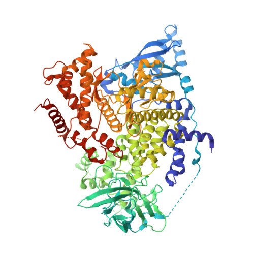
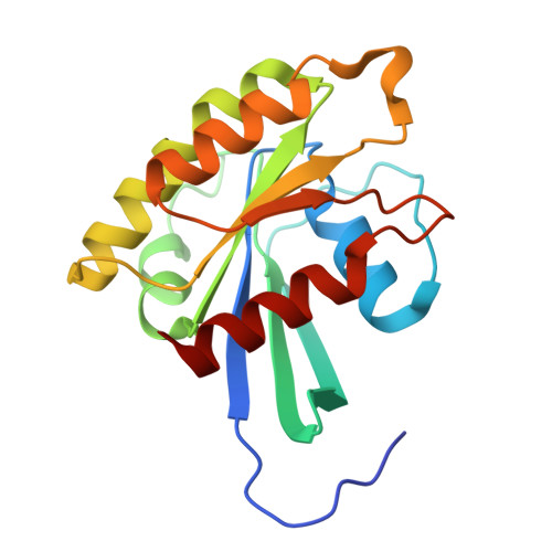
Structural insights into isoform-specific RAS-PI3K alpha interactions and the role of RAS in PI3K alpha activation.

Czyzyk, D.Yan, W.Messing, S.Gillette, W.Tsuji, T.Yamaguchi, M.Furuzono, S.Turner, D.M.Esposito, D.Nissley, D.V.McCormick, F.Simanshu, D.K.

(2025) Nat Commun 16: 525-525

  • DOI: https://doi.org/10.1038/s41467-024-55766-x
  • Primary Citation Related Structures: 
    9B4Q, 9B4R, 9B4S, 9B4T, 9C15

  • PubMed Abstract: 

    Mutations in RAS and PI3Kα are major drivers of human cancer. Their interaction plays a crucial role in activating PI3Kα and amplifying the PI3K-AKT-mTOR pathway. Disrupting RAS-PI3Kα interaction enhances survival in lung and skin cancer models and reduces tumor growth and angiogenesis, although the structural details of this interaction remain unclear. Here, we present structures of KRAS, RRAS2, and MRAS bound to the catalytic subunit (p110α) of PI3Kα, elucidating the interaction interfaces and local conformational changes upon complex formation. Structural and mutational analyses highlighted key residues in RAS and PI3Kα impacting binding affinity and revealed isoform-specific differences at the interaction interface in RAS and PI3K isoforms, providing a rationale for their differential affinities. Notably, in the RAS-p110α complex structures, RAS interaction with p110α is limited to the RAS-binding domain and does not involve the kinase domain. This study underscores the pivotal role of the RAS-PI3Kα interaction in PI3Kα activation and provides a blueprint for designing PI3Kα isoform-specific inhibitors to disrupt this interaction.


  • Organizational Affiliation
    • NCI RAS Initiative, Cancer Research Technology Program, Frederick National Laboratory for Cancer Research, Frederick, MD, USA.

Macromolecule Content 

  • Total Structure Weight: 133.35 kDa 
  • Atom Count: 8,714 
  • Modeled Residue Count: 1,063 
  • Deposited Residue Count: 1,144 
  • Unique protein chains: 2

Macromolecules

Find similar proteins by:|  3D Structure
Entity ID: 1
MoleculeChains  Sequence LengthOrganismDetailsImage
Phosphatidylinositol 4,5-bisphosphate 3-kinase catalytic subunit alpha isoform965Homo sapiensMutation(s): 3 
Gene Names: PIK3CA
EC: 2.7.1.137 (PDB Primary Data), 2.7.1.153 (PDB Primary Data), 2.7.11.1 (PDB Primary Data)
UniProt & NIH Common Fund Data Resources
Find proteins for P42336 (Homo sapiens)
Explore P42336 
Go to UniProtKB:  P42336
PHAROS:  P42336
GTEx:  ENSG00000121879 
Entity Groups
Sequence Clusters30% Identity50% Identity70% Identity90% Identity95% Identity100% Identity
UniProt GroupP42336
Sequence Annotations
Expand
Reference Sequence
Find similar proteins by:|  3D Structure
Entity ID: 2
MoleculeChains  Sequence LengthOrganismDetailsImage
Ras-related protein M-Ras179Homo sapiensMutation(s): 1 
Gene Names: MRASRRAS3
EC: 3.6.5.2
UniProt & NIH Common Fund Data Resources
Find proteins for O14807 (Homo sapiens)
Explore O14807 
Go to UniProtKB:  O14807
PHAROS:  O14807
GTEx:  ENSG00000158186 
Entity Groups
Sequence Clusters30% Identity50% Identity70% Identity90% Identity95% Identity100% Identity
UniProt GroupO14807
Sequence Annotations
Expand
Reference Sequence

Small Molecules

Ligands 3 Unique
IDChains Name / Formula / InChI Key2D Diagram3D Interactions
GNP
(Subject of Investigation/LOI)

Query on GNP


Download:Ideal Coordinates CCD File
D [auth B]PHOSPHOAMINOPHOSPHONIC ACID-GUANYLATE ESTER
C10 H17 N6 O13 P3
UQABYHGXWYXDTK-UUOKFMHZSA-N
5H5
(Subject of Investigation/LOI)

Query on 5H5


Download:Ideal Coordinates CCD File
C [auth A](2S)-2-({2-[1-(propan-2-yl)-1H-1,2,4-triazol-5-yl]-5,6-dihydroimidazo[1,2-d][1,4]benzoxazepin-9-yl}oxy)propanamide
C19 H22 N6 O3
SIKYDKLGPWRPMZ-LBPRGKRZSA-N
MG

Query on MG


Download:Ideal Coordinates CCD File
E [auth B]MAGNESIUM ION
Mg
JLVVSXFLKOJNIY-UHFFFAOYSA-N

Experimental Data & Validation

Experimental Data

  • Method: X-RAY DIFFRACTION
  • Resolution: 2.75 Å
  • R-Value Free:  0.262 (Depositor), 0.262 (DCC) 
  • R-Value Work:  0.213 (Depositor), 0.215 (DCC) 
  • R-Value Observed: 0.216 (Depositor) 
Space Group: P 1 21 1
Unit Cell:
Length ( Å )Angle ( ˚ )
a = 58.935α = 90
b = 105.936β = 97.5
c = 114.493γ = 90
Software Package:
Software NamePurpose
PHENIXrefinement
XDSdata scaling
XDSdata reduction
PHASERphasing

Structure Validation

View Full Validation Report


Ligand Structure Quality Assessment 


Entry History 

& Funding Information

Deposition Data


Funding OrganizationLocationGrant Number
National Institutes of Health/National Cancer Institute (NIH/NCI)United States75N91019D00024

Revision History  (Full details and data files)

  • Version 1.0: 2025-01-22
    Type: Initial release