9B4F | pdb_00009b4f

Structure of human PSS1-P269S


Experimental Data Snapshot

  • Method: ELECTRON MICROSCOPY
  • Resolution: 3.27 Å
  • Aggregation State: PARTICLE 
  • Reconstruction Method: SINGLE PARTICLE 

wwPDB Validation 3D Report Full Report

Validation slider image for 9B4F

This is version 1.3 of the entry. See complete history

Literature

Molecular insights into human phosphatidylserine synthase 1 reveal its inhibition promotes LDL uptake.

Long, T.Li, D.Vale, G.Jiang, Y.Schmiege, P.Yang, Z.J.McDonald, J.G.Li, X.

(2024) Cell 187: 5665-5678.e18

  • DOI: https://doi.org/10.1016/j.cell.2024.08.004
  • Primary Citation Related Structures: 
    9B4E, 9B4F, 9B4G

  • PubMed Abstract: 

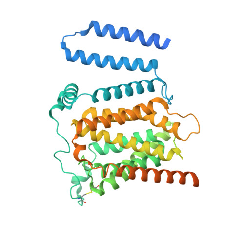
    In mammalian cells, two phosphatidylserine (PS) synthases drive PS synthesis. Gain-of-function mutations in the Ptdss1 gene lead to heightened PS production, causing Lenz-Majewski syndrome (LMS). Recently, pharmacological inhibition of PSS1 has been shown to suppress tumorigenesis. Here, we report the cryo-EM structures of wild-type human PSS1 (PSS1 WT ), the LMS-causing Pro269Ser mutant (PSS1 P269S ), and PSS1 WT in complex with its inhibitor DS55980254. PSS1 contains 10 transmembrane helices (TMs), with TMs 4-8 forming a catalytic core in the luminal leaflet. These structures revealed a working mechanism of PSS1 akin to the postulated mechanisms of the membrane-bound O-acyltransferase family. Additionally, we showed that both PS and DS55980254 can allosterically inhibit PSS1 and that inhibition by DS55980254 activates the SREBP pathways, thus enhancing the expression of LDL receptors and increasing cellular LDL uptake. This work uncovers a mechanism of mammalian PS synthesis and suggests that selective PSS1 inhibitors have the potential to lower blood cholesterol levels.


  • Organizational Affiliation
    • Department of Molecular Genetics, University of Texas Southwestern Medical Center, Dallas, TX 75390, USA.

Macromolecule Content 

  • Total Structure Weight: 113.23 kDa 
  • Atom Count: 5,612 
  • Modeled Residue Count: 670 
  • Deposited Residue Count: 962 
  • Unique protein chains: 1

Macromolecules

Find similar proteins by:|  3D Structure
Entity ID: 1
MoleculeChains  Sequence LengthOrganismDetailsImage
Phosphatidylserine synthase 1
A, B
481Homo sapiensMutation(s): 1 
Gene Names: PTDSS1KIAA0024PSSA
EC: 2.7.8.29
Membrane Entity: Yes 
UniProt & NIH Common Fund Data Resources
Find proteins for P48651 (Homo sapiens)
Explore P48651 
Go to UniProtKB:  P48651
PHAROS:  P48651
GTEx:  ENSG00000156471 
Entity Groups
Sequence Clusters30% Identity50% Identity70% Identity90% Identity95% Identity100% Identity
UniProt GroupP48651
Sequence Annotations
Expand
Reference Sequence

Experimental Data & Validation

Experimental Data

  • Method: ELECTRON MICROSCOPY
  • Resolution: 3.27 Å
  • Aggregation State: PARTICLE 
  • Reconstruction Method: SINGLE PARTICLE 
EM Software:
TaskSoftware PackageVersion
MODEL REFINEMENTPHENIX1.17_3644:

Structure Validation

View Full Validation Report


Entry History 

& Funding Information

Deposition Data


Funding OrganizationLocationGrant Number
National Institutes of Health/National Institute of General Medical Sciences (NIH/NIGMS)United StatesGM135343

Revision History  (Full details and data files)

  • Version 1.0: 2024-09-04
    Type: Initial release
  • Version 1.1: 2024-09-11
    Changes: Data collection, Database references
  • Version 1.2: 2024-10-16
    Changes: Data collection, Database references, Structure summary
  • Version 1.3: 2024-10-23
    Changes: Data collection