


Structure of human cardiac actin
Doran, M.H., Rynkiewicz, M.J., Sousa, D., Cammarato, A., Lehman, W.To be published.
Experimental Data Snapshot
wwPDB Validation 3D Report Full Report

Entity ID: 1 | |||||
|---|---|---|---|---|---|
| Molecule | Chains | Sequence Length | Organism | Details | Image |
| Actin, alpha cardiac muscle 1 | A, B [auth D], C [auth E] | 377 | Homo sapiens | Mutation(s): 1 Gene Names: ACTC1, ACTC EC: 3.6.4 | 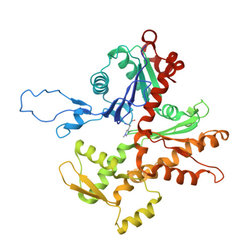 |
UniProt & NIH Common Fund Data Resources | |||||
PHAROS: P68032 GTEx: ENSG00000159251 | |||||
Entity Groups | |||||
| Sequence Clusters | 30% Identity50% Identity70% Identity90% Identity95% Identity100% Identity | ||||
| UniProt Group | P68032 | ||||
Sequence AnnotationsExpand | |||||
Reference Sequence | |||||
| Ligands 2 Unique | |||||
|---|---|---|---|---|---|
| ID | Chains | Name / Formula / InChI Key | 2D Diagram | 3D Interactions | |
| ADP Download:Ideal Coordinates CCD File | E [auth A], G [auth D], I [auth E] | ADENOSINE-5'-DIPHOSPHATE C10 H15 N5 O10 P2 XTWYTFMLZFPYCI-KQYNXXCUSA-N |  | ||
| MG Download:Ideal Coordinates CCD File | D [auth A], F [auth D], H [auth E] | MAGNESIUM ION Mg JLVVSXFLKOJNIY-UHFFFAOYSA-N |  | ||
| Modified Residues 1 Unique | |||||
|---|---|---|---|---|---|
| ID | Chains | Type | Formula | 2D Diagram | Parent |
| HIC Query on HIC | A, B [auth D], C [auth E] | L-PEPTIDE LINKING | C7 H11 N3 O2 |  | HIS |
| Task | Software Package | Version |
|---|---|---|
| MODEL REFINEMENT | PHENIX | |
| Funding Organization | Location | Grant Number |
|---|---|---|
| National Institutes of Health/National Heart, Lung, and Blood Institute (NIH/NHLBI) | United States | 1R56HL124091-01 |
| National Institutes of Health/National Heart, Lung, and Blood Institute (NIH/NHLBI) | United States | 5R01HL036153-33 |


