9B1F

Human urate transporter 1 URAT1 in apo state

  • Classification: MEMBRANE PROTEIN
  • Organism(s): Homo sapiens
  • Expression System: Homo sapiens
  • Mutation(s): No 
  • Membrane Protein: Yes  PDBTM

  • Deposited: 2024-03-13 Released: 2024-09-18 
  • Deposition Author(s): Dai, Y., Lee, C.H.
  • Funding Organization(s): National Institutes of Health/National Institute of General Medical Sciences (NIH/NIGMS)

Experimental Data Snapshot

  • Method: ELECTRON MICROSCOPY
  • Resolution: 2.90 Å
  • Aggregation State: PARTICLE 
  • Reconstruction Method: SINGLE PARTICLE 

wwPDB Validation   3D Report Full Report


This is version 1.0 of the entry. See complete history


Literature

Transport mechanism and structural pharmacology of human urate transporter URAT1.

Dai, Y.Lee, C.H.

(2024) Cell Res 

  • DOI: https://doi.org/10.1038/s41422-024-01023-1
  • Primary Citation of Related Structures:  
    9B1F, 9B1G, 9B1H, 9B1I, 9B1J, 9B1K, 9B1L, 9B1M, 9B1N, 9B1O

  • PubMed Abstract: 

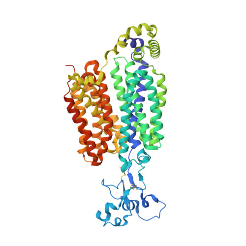
    Urate is an endogenous product of purine metabolism in the liver. High urate levels in the blood lead to gout, a very common and painful inflammatory arthritis. Excreted urate is reabsorbed in the kidney mainly by URAT1 antiporter, a key target for anti-gout drugs. To uncover the mechanisms of urate transport and drug inhibition, we determined cryo-EM structures of human URAT1 with urate, counter anion pyrazinoate, or anti-gout drugs of different chemotypes - lesinurad, verinurad, and dotinurad. We captured the outward-to-inward transition of URAT1 during urate uptake, revealing that urate binds in a phenylalanine-rich pocket and engages with key gating residues to drive the transport cycle. In contrast to the single binding site for urate, pyrazinoate interacts with three distinct, functionally relevant sites within URAT1, a mechanism that has not yet been observed in other anion antiporters. In addition, we found that while all three drugs compete with substrates and halt the transport cycle, verinurad and dotinurad further hijack gating residues to achieve high potency. These insights advance our understanding of organic anion transport and provide a foundation for designing improved gout therapeutics.


  • Organizational Affiliation

    Department of Structural Biology, St. Jude Children's Research Hospital, Memphis, TN, USA.


Macromolecules
Find similar proteins by:  (by identity cutoff)  |  3D Structure
Entity ID: 1
MoleculeChains Sequence LengthOrganismDetailsImage
Solute carrier family 22 member 12547Homo sapiensMutation(s): 0 
Gene Names: SLC22A12OATL4URAT1UNQ6453/PRO34004
Membrane Entity: Yes 
UniProt & NIH Common Fund Data Resources
Find proteins for Q96S37 (Homo sapiens)
Explore Q96S37 
Go to UniProtKB:  Q96S37
PHAROS:  Q96S37
GTEx:  ENSG00000197891 
Entity Groups  
Sequence Clusters30% Identity50% Identity70% Identity90% Identity95% Identity100% Identity
UniProt GroupQ96S37
Glycosylation
Glycosylation Sites: 1Go to GlyGen: Q96S37-1
Sequence Annotations
Expand
  • Reference Sequence
Small Molecules
Ligands 1 Unique
IDChains Name / Formula / InChI Key2D Diagram3D Interactions
NAG
Query on NAG

Download Ideal Coordinates CCD File 
B [auth A]2-acetamido-2-deoxy-beta-D-glucopyranose
C8 H15 N O6
OVRNDRQMDRJTHS-FMDGEEDCSA-N
Experimental Data & Validation

Experimental Data

  • Method: ELECTRON MICROSCOPY
  • Resolution: 2.90 Å
  • Aggregation State: PARTICLE 
  • Reconstruction Method: SINGLE PARTICLE 
EM Software:
TaskSoftware PackageVersion
MODEL REFINEMENTPHENIX1.21_5207:

Structure Validation

View Full Validation Report


Entry History & Funding Information

Deposition Data


Funding OrganizationLocationGrant Number
National Institutes of Health/National Institute of General Medical Sciences (NIH/NIGMS)United StatesGM143282

Revision History  (Full details and data files)

  • Version 1.0: 2024-09-18
    Type: Initial release