


Linkage and substrate specificity conferred by NZF ubiquitin binding domains
Michel, M.A., Scutts, S., Komander, D.To be published.
Experimental Data Snapshot
Starting Model: experimental
View more details
wwPDB Validation 3D Report Full Report

Entity ID: 1 | |||||
|---|---|---|---|---|---|
| Molecule | Chains | Sequence Length | Organism | Details | Image |
| Optineurin | A, B, D [auth C], E [auth D] | 96 | Homo sapiens | Mutation(s): 2 Gene Names: OPTN, FIP2, GLC1E, HIP7, HYPL, NRP |  |
UniProt & NIH Common Fund Data Resources | |||||
PHAROS: Q96CV9 GTEx: ENSG00000123240 | |||||
Entity Groups | |||||
| Sequence Clusters | 30% Identity50% Identity70% Identity90% Identity95% Identity100% Identity | ||||
| UniProt Group | Q96CV9 | ||||
Sequence AnnotationsExpand | |||||
Reference Sequence | |||||
Entity ID: 2 | |||||
|---|---|---|---|---|---|
| Molecule | Chains | Sequence Length | Organism | Details | Image |
| E3 ubiquitin-protein ligase RNF31 | C [auth G], F [auth E] | 30 | Homo sapiens | Mutation(s): 0 Gene Names: RNF31, ZIBRA EC: 2.3.2.31 | 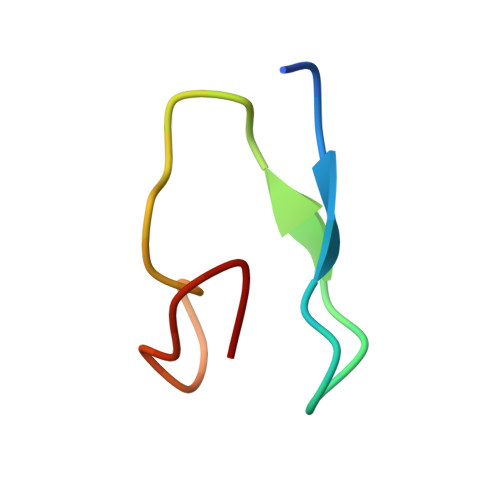 |
UniProt & NIH Common Fund Data Resources | |||||
PHAROS: Q96EP0 GTEx: ENSG00000092098 | |||||
Entity Groups | |||||
| Sequence Clusters | 30% Identity50% Identity70% Identity90% Identity95% Identity100% Identity | ||||
| UniProt Group | Q96EP0 | ||||
Sequence AnnotationsExpand | |||||
Reference Sequence | |||||
| Ligands 3 Unique | |||||
|---|---|---|---|---|---|
| ID | Chains | Name / Formula / InChI Key | 2D Diagram | 3D Interactions | |
| PG4 Download:Ideal Coordinates CCD File | I [auth B], N [auth C] | TETRAETHYLENE GLYCOL C8 H18 O5 UWHCKJMYHZGTIT-UHFFFAOYSA-N |  | ||
| PGE Download:Ideal Coordinates CCD File | G [auth A] H [auth B] J [auth B] K [auth B] M [auth G] | TRIETHYLENE GLYCOL C6 H14 O4 ZIBGPFATKBEMQZ-UHFFFAOYSA-N |  | ||
| ZN Download:Ideal Coordinates CCD File | L [auth G], S [auth E] | ZINC ION Zn PTFCDOFLOPIGGS-UHFFFAOYSA-N |  | ||
| Length ( Å ) | Angle ( ˚ ) |
|---|---|
| a = 49.755 | α = 90 |
| b = 92.684 | β = 90 |
| c = 149.84 | γ = 90 |
| Software Name | Purpose |
|---|---|
| PHENIX | refinement |
| Aimless | data scaling |
| XDS | data reduction |
| PHASER | phasing |
| Funding Organization | Location | Grant Number |
|---|---|---|
| Medical Research Council (MRC, United Kingdom) | United Kingdom | U105192732 |
| European Research Council (ERC) | European Union | 724804 |
| National Health and Medical Research Council (NHMRC, Australia) | Australia | GNT1178122 |


