9AXL

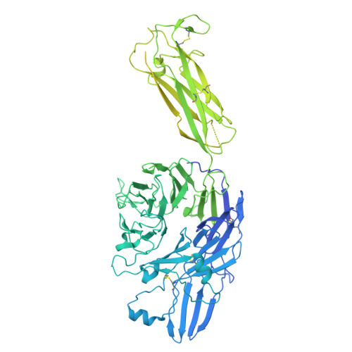
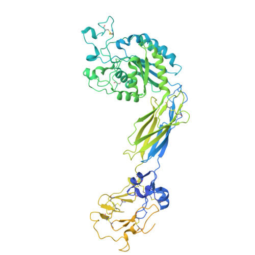
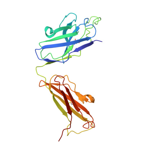
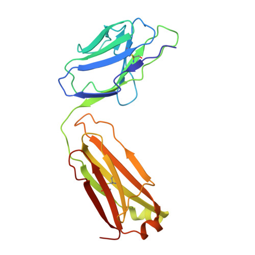
Structure of the semi-extended AlphaIIbBeta3 in complex with R21D10 Fab


Experimental Data Snapshot

  • Method: ELECTRON MICROSCOPY
  • Resolution: 3.30 Å
  • Aggregation State: PARTICLE 
  • Reconstruction Method: SINGLE PARTICLE 

Starting Models: experimental
View more details

wwPDB Validation   3D Report Full Report


This is version 1.1 of the entry. See complete history


Literature

An alpha IIb beta 3 monoclonal antibody traps a semiextended conformation and allosterically inhibits large ligand binding.

Wang, L.Wang, J.Li, J.Walz, T.Coller, B.S.

(2024) Blood Adv 8: 4398-4409

  • DOI: https://doi.org/10.1182/bloodadvances.2024013177
  • Primary Citation of Related Structures:  
    9AXL

  • PubMed Abstract: 

    Monoclonal antibodies (mAbs) have provided valuable information regarding the structure and function of platelet αIIbβ3. Protein disulfide isomerase (PDI) has been implicated in αIIbβ3 activation and binds to thrombin-activated αIIbβ3. Using human platelets as the immunogen, we identified a new mAb (R21D10) that inhibits the binding of PDI to platelets activated with thrombin receptor-activating peptide (T6). R21D10 also partially inhibited T6-induced fibrinogen and PAC-1 binding to platelets, as well as T6- and adenosine 5'-diphosphate-induced platelet aggregation. Mutual competition experiments showed that R21D10 does not inhibit the binding of mAbs 10E5 (anti-αIIb cap domain) or 7E3 (anti-β3 β-I domain), and immunoblot studies indicated that R21D10 binds to β3. The dissociation of αIIbβ3 by EDTA had a minimal effect on R21D10 binding. Cryogenic electron microscopy of the αIIbβ3-R21D10 Fab complex revealed that R21D10 binds to the β3 integrin-epidermal growth factor 1 (I-EGF1) domain and traps an intermediate conformation of αIIbβ3 with semiextended leg domains. The binding of R21D10 produces a major structural change in the β3 I-EGF2 domain associated with a new interaction between the β3 I-EGF2 and αIIb thigh domains, which may prevent the swing-out motion of the β3 hybrid domain required for high-affinity ligand binding and protect αIIbβ3 from EDTA-induced dissociation. R21D10 partially reversed the ligand binding priming effect of eptifibatide, suggesting that it could convert the swung-out conformation into a semiextended conformation. We concluded that R21D10 inhibits ligand binding to αIIbβ3 via a unique allosteric mechanism, which may or may not be related to its inhibition of PDI binding.


  • Organizational Affiliation

    Laboratory of Blood and Vascular Biology, The Rockefeller University, New York, NY.


Macromolecules
Find similar proteins by:  (by identity cutoff)  |  3D Structure
Entity ID: 1
MoleculeChains Sequence LengthOrganismDetailsImage
Integrin alpha-IIb1,039Homo sapiensMutation(s): 0 
UniProt & NIH Common Fund Data Resources
Find proteins for P08514 (Homo sapiens)
Explore P08514 
Go to UniProtKB:  P08514
PHAROS:  P08514
GTEx:  ENSG00000005961 
Entity Groups  
Sequence Clusters30% Identity50% Identity70% Identity90% Identity95% Identity100% Identity
UniProt GroupP08514
Sequence Annotations
Expand
  • Reference Sequence
Find similar proteins by:  (by identity cutoff)  |  3D Structure
Entity ID: 2
MoleculeChains Sequence LengthOrganismDetailsImage
Integrin beta-3788Homo sapiensMutation(s): 0 
UniProt & NIH Common Fund Data Resources
Find proteins for P05106 (Homo sapiens)
Explore P05106 
Go to UniProtKB:  P05106
PHAROS:  P05106
GTEx:  ENSG00000259207 
Entity Groups  
Sequence Clusters30% Identity50% Identity70% Identity90% Identity95% Identity100% Identity
UniProt GroupP05106
Sequence Annotations
Expand
  • Reference Sequence
Find similar proteins by:  (by identity cutoff)  |  3D Structure
Entity ID: 3
MoleculeChains Sequence LengthOrganismDetailsImage
R21D10 Fab heavy chainC [auth H]227Mus musculusMutation(s): 0 
Entity Groups  
Sequence Clusters30% Identity50% Identity70% Identity90% Identity95% Identity100% Identity
Sequence Annotations
Expand
  • Reference Sequence
Find similar proteins by:  (by identity cutoff)  |  3D Structure
Entity ID: 4
MoleculeChains Sequence LengthOrganismDetailsImage
R21D10 Fab light chainD [auth L]213Mus musculusMutation(s): 0 
Entity Groups  
Sequence Clusters30% Identity50% Identity70% Identity90% Identity95% Identity100% Identity
Sequence Annotations
Expand
  • Reference Sequence
Small Molecules
Ligands 2 Unique
IDChains Name / Formula / InChI Key2D Diagram3D Interactions
CA (Subject of Investigation/LOI)
Query on CA

Download Ideal Coordinates CCD File 
E [auth A]
F [auth A]
G [auth A]
H [auth A]
I [auth B]
E [auth A],
F [auth A],
G [auth A],
H [auth A],
I [auth B],
J [auth B]
CALCIUM ION
Ca
BHPQYMZQTOCNFJ-UHFFFAOYSA-N
MG (Subject of Investigation/LOI)
Query on MG

Download Ideal Coordinates CCD File 
K [auth B]MAGNESIUM ION
Mg
JLVVSXFLKOJNIY-UHFFFAOYSA-N
Experimental Data & Validation

Experimental Data

  • Method: ELECTRON MICROSCOPY
  • Resolution: 3.30 Å
  • Aggregation State: PARTICLE 
  • Reconstruction Method: SINGLE PARTICLE 
EM Software:
TaskSoftware PackageVersion
RECONSTRUCTIONcryoSPARCv4.4.0
MODEL REFINEMENTPHENIXversion 1.20.1-4487

Structure Validation

View Full Validation Report


Entry History & Funding Information

Deposition Data


Funding OrganizationLocationGrant Number
National Institutes of Health/National Heart, Lung, and Blood Institute (NIH/NHLBI)United States5R01HL019278-48

Revision History  (Full details and data files)

  • Version 1.0: 2024-11-13
    Type: Initial release
  • Version 1.1: 2024-11-27
    Changes: Data collection