9AU1 | pdb_00009au1

SARS-CoV-2 XBB.1.5 RBD bound to the VIR-7229 and the S309 Fab fragments


Experimental Data Snapshot

  • Method: X-RAY DIFFRACTION
  • Resolution: 2.41 Å
  • R-Value Free: 
    0.257 (Depositor), 0.256 (DCC) 
  • R-Value Work: 
    0.227 (Depositor), 0.225 (DCC) 
  • R-Value Observed: 
    0.228 (Depositor) 

Starting Model: experimental
View more details

wwPDB Validation 3D Report Full Report

Validation slider image for 9AU1

Ligand Structure Quality Assessment 


This is version 1.1 of the entry. See complete history

Literature

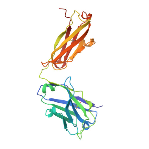
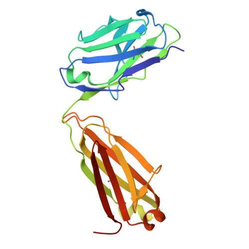
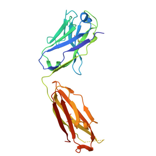
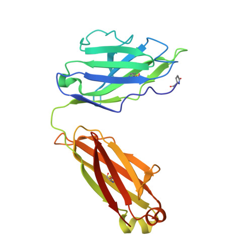
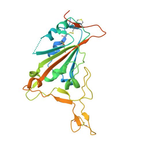
A potent pan-sarbecovirus neutralizing antibody resilient to epitope diversification.

Rosen, L.E.Tortorici, M.A.De Marco, A.Pinto, D.Foreman, W.B.Taylor, A.L.Park, Y.J.Bohan, D.Rietz, T.Errico, J.M.Hauser, K.Dang, H.V.Chartron, J.W.Giurdanella, M.Cusumano, G.Saliba, C.Zatta, F.Sprouse, K.R.Addetia, A.Zepeda, S.K.Brown, J.Lee, J.Dellota Jr., E.Rajesh, A.Noack, J.Tao, Q.DaCosta, Y.Tsu, B.Acosta, R.Subramanian, S.de Melo, G.D.Kergoat, L.Zhang, I.Liu, Z.Guarino, B.Schmid, M.A.Schnell, G.Miller, J.L.Lempp, F.A.Czudnochowski, N.Cameroni, E.Whelan, S.P.J.Bourhy, H.Purcell, L.A.Benigni, F.di Iulio, J.Pizzuto, M.S.Lanzavecchia, A.Telenti, A.Snell, G.Corti, D.Veesler, D.Starr, T.N.

(2024) Cell 187: 7196

  • DOI: https://doi.org/10.1016/j.cell.2024.09.026
  • Primary Citation Related Structures: 
    8S6M, 9ASD, 9ATM, 9AU1, 9AU2

  • PubMed Abstract: 

    Severe acute respiratory syndrome coronavirus 2 (SARS-CoV-2) evolution has resulted in viral escape from clinically authorized monoclonal antibodies (mAbs), creating a need for mAbs that are resilient to epitope diversification. Broadly neutralizing coronavirus mAbs that are sufficiently potent for clinical development and retain activity despite viral evolution remain elusive. We identified a human mAb, designated VIR-7229, which targets the viral receptor-binding motif (RBM) with unprecedented cross-reactivity to all sarbecovirus clades, including non-ACE2-utilizing bat sarbecoviruses, while potently neutralizing SARS-CoV-2 variants since 2019, including the recent EG.5, BA.2.86, and JN.1. VIR-7229 tolerates extraordinary epitope variability, partly attributed to its high binding affinity, receptor molecular mimicry, and interactions with RBM backbone atoms. Consequently, VIR-7229 features a high barrier for selection of escape mutants, which are rare and associated with reduced viral fitness, underscoring its potential to be resilient to future viral evolution. VIR-7229 is a strong candidate to become a next-generation medicine.


  • Organizational Affiliation
    • Vir Biotechnology, San Francisco, CA 94158, USA.

Macromolecule Content 

  • Total Structure Weight: 124.04 kDa 
  • Atom Count: 8,190 
  • Modeled Residue Count: 1,066 
  • Deposited Residue Count: 1,133 
  • Unique protein chains: 5

Macromolecules

Find similar proteins by:|  3D Structure
Entity ID: 1
MoleculeChains  Sequence LengthOrganismDetailsImage
S309 Fab heavy chainA [auth M]230Homo sapiensMutation(s): 0 
Find similar proteins by:|  3D Structure
Entity ID: 2
MoleculeChains  Sequence LengthOrganismDetailsImage
S309 Fab light chainB [auth N]212Homo sapiensMutation(s): 0 
Find similar proteins by:|  3D Structure
Entity ID: 3
MoleculeChains  Sequence LengthOrganismDetailsImage
VIR-7229 Fab heavy chainC [auth H]225Homo sapiensMutation(s): 0 
Find similar proteins by:|  3D Structure
Entity ID: 4
MoleculeChains  Sequence LengthOrganismDetailsImage
VIR-7229 Fab light chainD [auth L]216Homo sapiensMutation(s): 0 
Find similar proteins by:|  3D Structure
Entity ID: 5
MoleculeChains  Sequence LengthOrganismDetailsImage
SARS-CoV-2 XBB.1.5 RBDE [auth R]250Severe acute respiratory syndrome coronavirus 2Mutation(s): 0 
UniProt
Find proteins for A0A8A5XRG7 (Severe acute respiratory syndrome coronavirus 2)
Explore A0A8A5XRG7 
Go to UniProtKB:  A0A8A5XRG7
Entity Groups
Sequence Clusters30% Identity50% Identity70% Identity90% Identity95% Identity100% Identity
UniProt GroupA0A8A5XRG7
Glycosylation
Glycosylation Sites: 1
Sequence Annotations
Expand
Reference Sequence

Small Molecules

Ligands 5 Unique
IDChains Name / Formula / InChI Key2D Diagram3D Interactions
EPE

Query on EPE


Download:Ideal Coordinates CCD File
I [auth L]4-(2-HYDROXYETHYL)-1-PIPERAZINE ETHANESULFONIC ACID
C8 H18 N2 O4 S
JKMHFZQWWAIEOD-UHFFFAOYSA-N
NAG

Query on NAG


Download:Ideal Coordinates CCD File
K [auth R]2-acetamido-2-deoxy-beta-D-glucopyranose
C8 H15 N O6
OVRNDRQMDRJTHS-FMDGEEDCSA-N
EDO

Query on EDO


Download:Ideal Coordinates CCD File
G [auth H],
J [auth R]
1,2-ETHANEDIOL
C2 H6 O2
LYCAIKOWRPUZTN-UHFFFAOYSA-N
ACY

Query on ACY


Download:Ideal Coordinates CCD File
H [auth L]ACETIC ACID
C2 H4 O2
QTBSBXVTEAMEQO-UHFFFAOYSA-N
K

Query on K


Download:Ideal Coordinates CCD File
F [auth N]POTASSIUM ION
K
NPYPAHLBTDXSSS-UHFFFAOYSA-N
Modified Residues  1 Unique
IDChains TypeFormula2D DiagramParent
PCA
Query on PCA
D [auth L]L-PEPTIDE LINKINGC5 H7 N O3GLN

Experimental Data & Validation

Experimental Data

  • Method: X-RAY DIFFRACTION
  • Resolution: 2.41 Å
  • R-Value Free:  0.257 (Depositor), 0.256 (DCC) 
  • R-Value Work:  0.227 (Depositor), 0.225 (DCC) 
  • R-Value Observed: 0.228 (Depositor) 
Space Group: P 21 21 21
Unit Cell:
Length ( Å )Angle ( ˚ )
a = 73.726α = 90
b = 112.525β = 90
c = 170.314γ = 90
Software Package:
Software NamePurpose
PHENIXrefinement
PHASERphasing
XDSdata reduction
Aimlessdata scaling

Structure Validation

View Full Validation Report


Ligand Structure Quality Assessment 


Entry History 

& Funding Information

Deposition Data


Funding OrganizationLocationGrant Number
National Institutes of Health/National Institute Of Allergy and Infectious Diseases (NIH/NIAID)United States75N93022C00036

Revision History  (Full details and data files)

  • Version 1.0: 2024-10-16
    Type: Initial release
  • Version 1.1: 2024-12-25
    Changes: Database references