9ARF

Rat GluN1-GluN2B NMDA receptor channel in complex with glycine, glutamate, and EU-1622-A, in nonactive1 conformation

  • Classification: MEMBRANE PROTEIN
  • Organism(s): Rattus norvegicus
  • Expression System: Spodoptera frugiperda
  • Mutation(s): No 
  • Membrane Protein: Yes  PDBTM

  • Deposited: 2024-02-23 Released: 2024-07-31 
  • Deposition Author(s): Chou, T.-H., Furukawa, H.
  • Funding Organization(s): National Institutes of Health/National Institute of Neurological Disorders and Stroke (NIH/NINDS), National Institutes of Health/National Institute of Mental Health (NIH/NIMH)

Experimental Data Snapshot

  • Method: ELECTRON MICROSCOPY
  • Resolution: 3.13 Å
  • Aggregation State: PARTICLE 
  • Reconstruction Method: SINGLE PARTICLE 

Starting Model: experimental
View more details

wwPDB Validation   3D Report Full Report


This is version 1.2 of the entry. See complete history


Literature

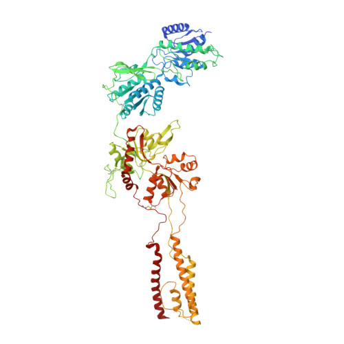
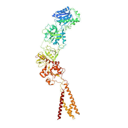
Molecular mechanism of ligand gating and opening of NMDA receptor.

Chou, T.H.Epstein, M.Fritzemeier, R.G.Akins, N.S.Paladugu, S.Ullman, E.Z.Liotta, D.C.Traynelis, S.F.Furukawa, H.

(2024) Nature 632: 209-217

  • DOI: https://doi.org/10.1038/s41586-024-07742-0
  • Primary Citation of Related Structures:  
    9ARE, 9ARF, 9ARG, 9ARH, 9ARI, 9BIB

  • PubMed Abstract: 

    Glutamate transmission and activation of ionotropic glutamate receptors are the fundamental means by which neurons control their excitability and neuroplasticity 1 . The N-methyl-D-aspartate receptor (NMDAR) is unique among all ligand-gated channels, requiring two ligands-glutamate and glycine-for activation. These receptors function as heterotetrameric ion channels, with the channel opening dependent on the simultaneous binding of glycine and glutamate to the extracellular ligand-binding domains (LBDs) of the GluN1 and GluN2 subunits, respectively 2,3 . The exact molecular mechanism for channel gating by the two ligands has been unclear, particularly without structures representing the open channel and apo states. Here we show that the channel gate opening requires tension in the linker connecting the LBD and transmembrane domain (TMD) and rotation of the extracellular domain relative to the TMD. Using electron cryomicroscopy, we captured the structure of the GluN1-GluN2B (GluN1-2B) NMDAR in its open state bound to a positive allosteric modulator. This process rotates and bends the pore-forming helices in GluN1 and GluN2B, altering the symmetry of the TMD channel from pseudofourfold to twofold. Structures of GluN1-2B NMDAR in apo and single-liganded states showed that binding of either glycine or glutamate alone leads to distinct GluN1-2B dimer arrangements but insufficient tension in the LBD-TMD linker for channel opening. This mechanistic framework identifies a key determinant for channel gating and a potential pharmacological strategy for modulating NMDAR activity.


  • Organizational Affiliation

    W.M. Keck Structural Biology Laboratory, Cold Spring Harbor Laboratory, Cold Spring Harbor, NY, USA.


Macromolecules
Find similar proteins by:  (by identity cutoff)  |  3D Structure
Entity ID: 1
MoleculeChains Sequence LengthOrganismDetailsImage
Glutamate receptor ionotropic, NMDA 1
A, C
847Rattus norvegicusMutation(s): 0 
Gene Names: Grin1Nmdar1
Membrane Entity: Yes 
UniProt
Find proteins for P35439 (Rattus norvegicus)
Explore P35439 
Go to UniProtKB:  P35439
Entity Groups  
Sequence Clusters30% Identity50% Identity70% Identity90% Identity95% Identity100% Identity
UniProt GroupP35439
Sequence Annotations
Expand
  • Reference Sequence
Find similar proteins by:  (by identity cutoff)  |  3D Structure
Entity ID: 2
MoleculeChains Sequence LengthOrganismDetailsImage
Glutamate receptor ionotropic, NMDA 2B
B, D
883Rattus norvegicusMutation(s): 0 
Gene Names: Grin2b
Membrane Entity: Yes 
UniProt
Find proteins for Q00960 (Rattus norvegicus)
Explore Q00960 
Go to UniProtKB:  Q00960
Entity Groups  
Sequence Clusters30% Identity50% Identity70% Identity90% Identity95% Identity100% Identity
UniProt GroupQ00960
Glycosylation
Glycosylation Sites: 2Go to GlyGen: Q00960-1
Sequence Annotations
Expand
  • Reference Sequence
Oligosaccharides

Help

Entity ID: 3
MoleculeChains Length2D Diagram Glycosylation3D Interactions
2-acetamido-2-deoxy-beta-D-glucopyranose-(1-4)-2-acetamido-2-deoxy-beta-D-glucopyranose
E, F
2N-Glycosylation
Glycosylation Resources
GlyTouCan:  G42666HT
GlyCosmos:  G42666HT
GlyGen:  G42666HT
Experimental Data & Validation

Experimental Data

  • Method: ELECTRON MICROSCOPY
  • Resolution: 3.13 Å
  • Aggregation State: PARTICLE 
  • Reconstruction Method: SINGLE PARTICLE 
EM Software:
TaskSoftware PackageVersion
RECONSTRUCTIONcryoSPARC
MODEL REFINEMENTPHENIX

Structure Validation

View Full Validation Report


Entry History & Funding Information

Deposition Data


Funding OrganizationLocationGrant Number
National Institutes of Health/National Institute of Neurological Disorders and Stroke (NIH/NINDS)United StatesNS111745
National Institutes of Health/National Institute of Mental Health (NIH/NIMH)United StatesMH085926

Revision History  (Full details and data files)

  • Version 1.0: 2024-07-31
    Type: Initial release
  • Version 1.1: 2024-08-07
    Changes: Data collection, Database references
  • Version 1.2: 2024-08-14
    Changes: Data collection, Database references