8ZY2 | pdb_00008zy2

Sarbecovirus BANAL-20-52 Spike Trimer in a Locked Conformation


Experimental Data Snapshot

  • Method: ELECTRON MICROSCOPY
  • Resolution: 2.90 Å
  • Aggregation State: PARTICLE 
  • Reconstruction Method: SINGLE PARTICLE 

wwPDB Validation 3D Report Full Report

Validation slider image for 8ZY2

This is version 1.4 of the entry. See complete history

Literature

SARS-related coronavirus S-protein structures reveal synergistic RBM interactions underpinning high-affinity human ACE2 binding.

Wang, J.Ma, Y.Li, Z.Yuan, H.Liu, B.Li, Z.Su, M.Habib, G.Liu, Y.Fu, L.Wang, P.Li, M.He, J.Chen, J.Zhou, P.Shi, Z.Chen, X.Xiong, X.

(2025) Sci Adv 11: eadr8772-eadr8772

  • DOI: https://doi.org/10.1126/sciadv.adr8772
  • Primary Citation Related Structures: 
    8ZY0, 8ZY1, 8ZY2, 8ZY3, 8ZY4, 8ZY5, 8ZY6, 8ZY7, 8ZY9, 8ZYA

  • PubMed Abstract: 

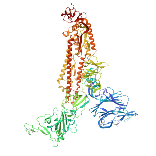
    High-affinity and specific binding toward the human angiotensin-converting enzyme 2 (hACE2) receptor by severe acute respiratory syndrome coronavirus (SARS)-related coronaviruses (SARSr-CoVs) remains incompletely understood. We report cryo-electron microscopy structures of eight different S-proteins from SARSr-CoVs found across Asia, Europe, and Africa. These S-proteins all adopt tightly packed, locked, prefusion conformations. These structures enable the classification of SARSr-CoV S-proteins into three types, based on their receptor-binding motif (RBM) structures and ACE2 binding characteristics. Type-2 S-proteins often preferentially bind bat ACE2 (bACE2) over hACE2. We report a structure of a type-2 BtKY72-RBD in complex with bACE2 to understand ACE2 specificity. Structure-guided mutagenesis of BtKY72-RBD reveals that multiple synergistic mutations in four different regions of RBM are required to achieve high-affinity hACE2 binding. Similar RBM changes can also confer hACE2 binding to another type-2 BM48-31 S-protein, which is primarily non-ACE2 binding. These results provide an understanding of how high-affinity hACE2 binding may be acquired by SARSr-CoV S-proteins.


  • Organizational Affiliation
    • Guangdong Provincial Key Laboratory of Stem Cell and Regenerative Medicine, Guangdong-Hong Kong Joint Research Laboratory for Stem Cell and Regenerative Medicine, Guangzhou Institutes of Biomedicine and Health, Chinese Academy of Sciences, Guangzhou, China.

Macromolecule Content 

  • Total Structure Weight: 396.04 kDa 
  • Atom Count: 27,099 
  • Modeled Residue Count: 3,309 
  • Deposited Residue Count: 3,408 
  • Unique protein chains: 1

Macromolecules

Find similar proteins by:|  3D Structure
Entity ID: 1
MoleculeChains  Sequence LengthOrganismDetailsImage
Spike glycoprotein
A, B, C
1,136SarbecovirusMutation(s): 0 
UniProt
Find proteins for P0DTC2 (Severe acute respiratory syndrome coronavirus 2)
Explore P0DTC2 
Go to UniProtKB:  P0DTC2
Entity Groups
Sequence Clusters30% Identity50% Identity70% Identity90% Identity95% Identity100% Identity
UniProt GroupP0DTC2
Glycosylation
Glycosylation Sites: 18Go to GlyGen: P0DTC2-1
Sequence Annotations
Expand
Reference Sequence

Small Molecules

Ligands 3 Unique
IDChains Name / Formula / InChI Key2D Diagram3D Interactions
BLA
(Subject of Investigation/LOI)

Query on BLA


Download:Ideal Coordinates CCD File
JA [auth A],
KB [auth C],
XA [auth B]
BILIVERDINE IX ALPHA
C33 H34 N4 O6
GWZYPXHJIZCRAJ-SRVCBVSDSA-N
EIC

Query on EIC


Download:Ideal Coordinates CCD File
KA [auth A],
LA [auth B],
YA [auth C]
LINOLEIC ACID
C18 H32 O2
OYHQOLUKZRVURQ-HZJYTTRNSA-N
NAG

Query on NAG


Download:Ideal Coordinates CCD File
AA [auth A]
AB [auth C]
BA [auth A]
BB [auth C]
CA [auth A]
AA [auth A],
AB [auth C],
BA [auth A],
BB [auth C],
CA [auth A],
CB [auth C],
DA [auth A],
DB [auth C],
EA [auth A],
EB [auth C],
FA [auth A],
FB [auth C],
GA [auth A],
GB [auth C],
HA [auth A],
HB [auth C],
IA [auth A],
IB [auth C],
JB [auth C],
MA [auth B],
NA [auth B],
OA [auth B],
PA [auth B],
QA [auth B],
RA [auth B],
SA [auth B],
TA [auth B],
UA [auth B],
VA [auth B],
WA [auth B],
Y [auth A],
Z [auth A],
ZA [auth C]
2-acetamido-2-deoxy-beta-D-glucopyranose
C8 H15 N O6
OVRNDRQMDRJTHS-FMDGEEDCSA-N

Experimental Data & Validation

Experimental Data

  • Method: ELECTRON MICROSCOPY
  • Resolution: 2.90 Å
  • Aggregation State: PARTICLE 
  • Reconstruction Method: SINGLE PARTICLE 
EM Software:
TaskSoftware PackageVersion
MODEL REFINEMENTPHENIX

Structure Validation

View Full Validation Report


Entry History 

& Funding Information

Deposition Data


Funding OrganizationLocationGrant Number
Other government2022A1515110495
Other governmentSRPG22-002

Revision History  (Full details and data files)

  • Version 1.0: 2025-02-05
    Type: Initial release
  • Version 1.1: 2025-02-12
    Changes: Data collection, Structure summary
  • Version 1.2: 2025-02-26
    Changes: Data collection, Structure summary
  • Version 1.3: 2025-06-18
    Changes: Data collection, Database references
  • Version 1.4: 2025-07-23
    Changes: Data collection