


Crystal structure of N terminal deletion mutant of Staphylococcal atypical two-cysteine thiol peroxidase complexed with substrate analog
Shukla, M., Maji, S., Yadav, V.K., Bhattacharyya, S.To be published.
Experimental Data Snapshot
Starting Model: experimental
View more details
Entity ID: 1 | |||||
|---|---|---|---|---|---|
| Molecule | Chains | Sequence Length | Organism | Details | Image |
| Thiol peroxidase | 149 | Staphylococcus aureus | Mutation(s): 0 Gene Names: tpx, SACOL1762 EC: 1.11.1.24 | 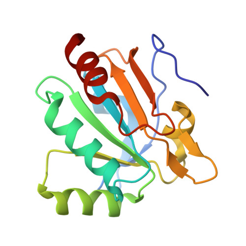 | |
UniProt | |||||
Entity Groups | |||||
| Sequence Clusters | 30% Identity50% Identity70% Identity90% Identity95% Identity100% Identity | ||||
| UniProt Group | Q5HF61 | ||||
Sequence AnnotationsExpand | |||||
Reference Sequence | |||||
| Ligands 6 Unique | |||||
|---|---|---|---|---|---|
| ID | Chains | Name / Formula / InChI Key | 2D Diagram | 3D Interactions | |
| PG4 Download:Ideal Coordinates CCD File | E [auth A] F [auth A] G [auth A] H [auth A] K [auth B] | TETRAETHYLENE GLYCOL C8 H18 O5 UWHCKJMYHZGTIT-UHFFFAOYSA-N |  | ||
| DTO Download:Ideal Coordinates CCD File | O [auth B] | 1-HYDROXYSULFANYL-4-MERCAPTO-BUTANE-2,3-DIOL C4 H10 O3 S2 MFLGZMZEMVZQCC-QWWZWVQMSA-N |  | ||
| DTU Download:Ideal Coordinates CCD File | U [auth D] | (2R,3S)-1,4-DIMERCAPTOBUTANE-2,3-DIOL C4 H10 O2 S2 VHJLVAABSRFDPM-ZXZARUISSA-N |  | ||
| A1L2G (Subject of Investigation/LOI) Download:Ideal Coordinates CCD File | J [auth A], R [auth C], S [auth D] | O-tert-Butylhydroxylamine C4 H11 N O KKVUFSINQFSJNK-UHFFFAOYSA-N |  | ||
| IMD Download:Ideal Coordinates CCD File | N [auth B] | IMIDAZOLE C3 H5 N2 RAXXELZNTBOGNW-UHFFFAOYSA-O |  | ||
| PEO Download:Ideal Coordinates CCD File | I [auth A], P [auth B], Q [auth B] | HYDROGEN PEROXIDE H2 O2 MHAJPDPJQMAIIY-UHFFFAOYSA-N |  | ||
| Length ( Å ) | Angle ( ˚ ) |
|---|---|
| a = 38.267 | α = 90 |
| b = 148.331 | β = 90 |
| c = 72.952 | γ = 90 |
| Software Name | Purpose |
|---|---|
| REFMAC | refinement |
| SCALA | data scaling |
| XDS | data reduction |
| PHASER | phasing |
| Funding Organization | Location | Grant Number |
|---|---|---|
| Science and Engineering Research Board (SERB) | India | SRG/2020/001353 |


