8ZUH

Crystal structure of bovine Fbs2/Skp1/Man3GlcNAc2 complex


Experimental Data Snapshot

  • Method: X-RAY DIFFRACTION
  • Resolution: 3.20 Å
  • R-Value Free: 0.247 
  • R-Value Work: 0.206 
  • R-Value Observed: 0.208 

Starting Models: in silico, experimental
View more details

wwPDB Validation   3D Report Full Report


This is version 1.1 of the entry. See complete history


Literature

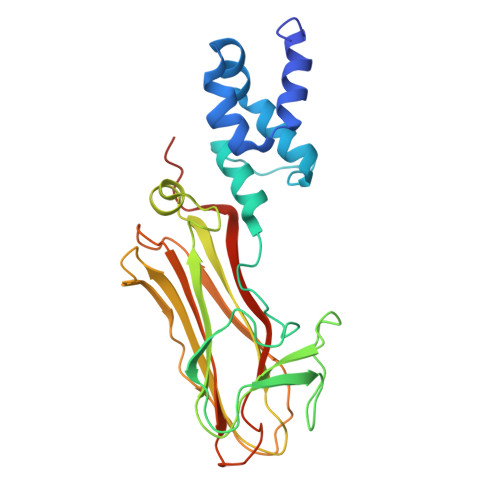
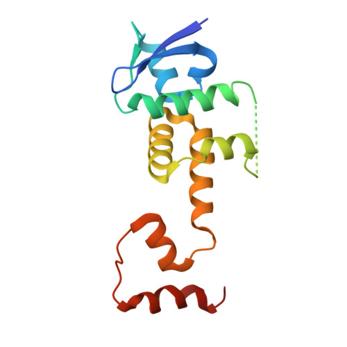
Structural basis of sugar recognition by SCF FBS2 ubiquitin ligase involved in NGLY1 deficiency.

Satoh, T.Yagi-Utsumi, M.Ishii, N.Mizushima, T.Yagi, H.Kato, R.Tachida, Y.Tateno, H.Matsuo, I.Kato, K.Suzuki, T.Yoshida, Y.

(2024) FEBS Lett 598: 2259-2268

  • DOI: https://doi.org/10.1002/1873-3468.15003
  • Primary Citation of Related Structures:  
    8ZUH

  • PubMed Abstract: 

    The cytosolic peptide:N-glycanase (PNGase) is involved in the quality control of N-glycoproteins via the endoplasmic reticulum-associated degradation (ERAD) pathway. Mutations in the gene encoding cytosolic PNGase (NGLY1 in humans) cause NGLY1 deficiency. Recent findings indicate that the F-box protein FBS2 of the SCF FBS2 ubiquitin ligase complex can be a promising drug target for NGLY1 deficiency. Here, we determined the crystal structure of bovine FBS2 complexed with the adaptor protein SKP1 and a sugar ligand, Man 3 GlcNAc 2 , which corresponds to the core pentasaccharide of N-glycan. Our crystallographic data together with NMR data revealed the structural basis of disparate sugar-binding specificities in homologous FBS proteins and identified a potential druggable pocket for in silico docking studies. Our results provide a potential basis for the development of selective inhibitors against FBS2 in NGLY1 deficiency.


  • Organizational Affiliation

    Graduate School of Pharmaceutical Sciences, Nagoya City University, Japan.


Macromolecules
Find similar proteins by:  (by identity cutoff)  |  3D Structure
Entity ID: 1
MoleculeChains Sequence LengthOrganismDetailsImage
F-box only protein 6
A, C
265Bos taurusMutation(s): 0 
Gene Names: FBXO6FBS2FBX6
UniProt
Find proteins for Q3SX24 (Bos taurus)
Explore Q3SX24 
Go to UniProtKB:  Q3SX24
Entity Groups  
Sequence Clusters30% Identity50% Identity70% Identity90% Identity95% Identity100% Identity
UniProt GroupQ3SX24
Sequence Annotations
Expand
  • Reference Sequence
Find similar proteins by:  (by identity cutoff)  |  3D Structure
Entity ID: 2
MoleculeChains Sequence LengthOrganismDetailsImage
S-phase kinase-associated protein 1166Bos taurusMutation(s): 0 
Gene Names: SKP1SKP1A
UniProt
Find proteins for Q3ZCF3 (Bos taurus)
Explore Q3ZCF3 
Go to UniProtKB:  Q3ZCF3
Entity Groups  
Sequence Clusters30% Identity50% Identity70% Identity90% Identity95% Identity100% Identity
UniProt GroupQ3ZCF3
Sequence Annotations
Expand
  • Reference Sequence
Oligosaccharides

Help

Entity ID: 3
MoleculeChains Length2D Diagram Glycosylation3D Interactions
alpha-D-mannopyranose-(1-3)-[alpha-D-mannopyranose-(1-6)]beta-D-mannopyranose-(1-4)-2-acetamido-2-deoxy-beta-D-glucopyranose-(1-4)-2-acetamido-2-deoxy-beta-D-glucopyranose
D, E
5N/A
Glycosylation Resources
GlyTouCan:  G22768VO
GlyCosmos:  G22768VO
GlyGen:  G22768VO
Experimental Data & Validation

Experimental Data

  • Method: X-RAY DIFFRACTION
  • Resolution: 3.20 Å
  • R-Value Free: 0.247 
  • R-Value Work: 0.206 
  • R-Value Observed: 0.208 
  • Space Group: P 41
Unit Cell:
Length ( Å )Angle ( ˚ )
a = 115.72α = 90
b = 115.72β = 90
c = 110.384γ = 90
Software Package:
Software NamePurpose
PHENIXrefinement
XDSdata reduction
XDSdata scaling
MOLREPphasing

Structure Validation

View Full Validation Report


Entry History & Funding Information

Deposition Data


Funding OrganizationLocationGrant Number
Japan Agency for Medical Research and Development (AMED)JapanJP24gm1410003

Revision History  (Full details and data files)

  • Version 1.0: 2024-09-04
    Type: Initial release
  • Version 1.1: 2024-10-09
    Changes: Database references, Structure summary