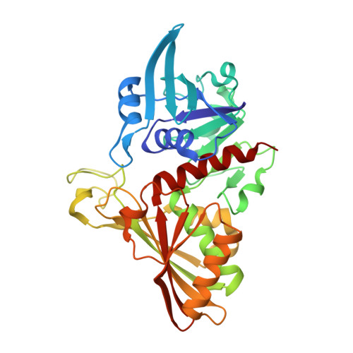
Crystal structure of the complex of glyceraldehyde-3-phosphate dehydrogenase of type B from Acinetobacter baumannii with Adenosine monophosphate at 3.20 A resolution.
Pahuja, P., Viswanathan, V., Kumari, A., Singh, A., Kumar, A., Sharma, P., Chopra, S., Sharma, S., Raje, C.I., Singh, T.P.To be published.