A red fluorescent genetically encoded biosensor for in vivo imaging of extracellular L-lactate dynamics.
Kamijo, Y., Machler, P., Ness, N., Vu, C.Q., Kusakizako, T., Mannuthodikayil, J., Ku, Z., Boisvert, M., Grebenik, E., Miyazaki, I., Hashizume, R., Sato, H., Liu, R., Hori, Y., Tomita, T., Katayama, T., Furube, A., Caraveo, G., Paquet, M.E., Drobizhev, M., Nureki, O., Arai, S., Brancaccio, M., Campbell, R.E., Kleinfeld, D., Nasu, Y.(2025) Nat Commun 16: 9531-9531
- PubMed: 41162396 Search on PubMedSearch on PubMed Central
- DOI: https://doi.org/10.1038/s41467-025-64484-x
- Primary Citation Related Structures:
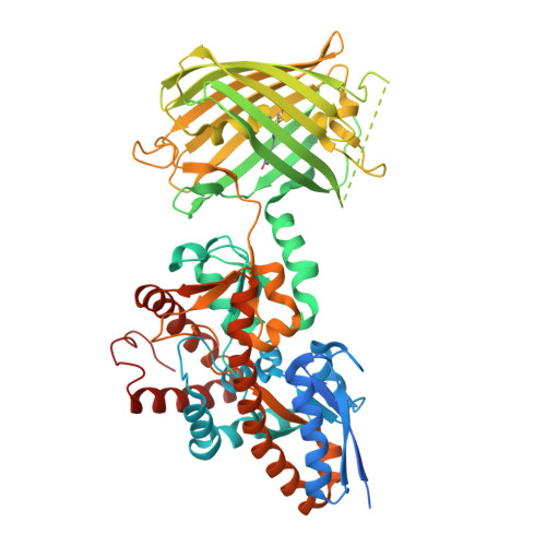
8ZMZ - PubMed Abstract:
L-Lactate is increasingly recognized as an intercellular energy currency in mammals, but mysteries remain regarding the spatial and temporal dynamics of its release and uptake between cells via the extracellular environment. Here we introduce R-eLACCO2.1, a red fluorescent extracellular L-lactate biosensor that is superior to previously reported green fluorescent biosensors in in vivo sensitivity to increases in extracellular L-lactate and spectral orthogonality. R-eLACCO2.1 exhibits excellent fluorescence response in cultured cells, mouse brain slices, and live mice. R-eLACCO2.1 also serves as an effective fluorescence lifetime-based biosensor. Using R-eLACCO2.1, we monitor whisker stimulation and locomotion-induced changes in endogenous extracellular L-lactate in the somatosensory cortex of awake mice. To highlight the potential insights gained from in vivo measurements with R-eLACCO2.1, we perform dual-color imaging from the somatosensory cortex of actively locomoting mice. This enables us to simultaneously observe the neural activity, reported by a green fluorescent GCaMP calcium ion biosensor, and extracellular L-lactate. As the high-performance tool in the suite of extracellular L-lactate biosensors, R-eLACCO2.1 is ideally suited to delimit the emerging roles of L-lactate in mammalian metabolism.
- Department of Chemistry, School of Science, The University of Tokyo, Bunkyo-ku, Tokyo, Japan.
Organizational Affiliation: