8ZKC

iron-sulfur cluster transfer protein ApbC


Experimental Data Snapshot

  • Method: X-RAY DIFFRACTION
  • Resolution: 2.85 Å
  • R-Value Free: 0.279 
  • R-Value Work: 0.235 
  • R-Value Observed: 0.237 

Starting Model: in silico
View more details

wwPDB Validation   3D Report Full Report


This is version 1.0 of the entry. See complete history


Literature

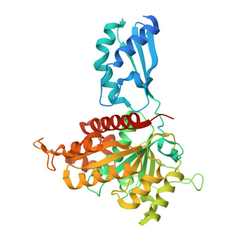
Crystal structure of the iron-sulfur cluster transfer protein ApbC from Escherichia coli.

Yang, J.Duan, Y.F.Liu, L.

(2024) Biochem Biophys Res Commun 722: 150167-150167

  • DOI: https://doi.org/10.1016/j.bbrc.2024.150167
  • Primary Citation of Related Structures:  
    8ZKC

  • PubMed Abstract: 

    Iron-sulfur (Fe-S) clusters are ubiquitous and are necessary to sustain basic life processes. The intracellular Fe-S clusters do not form spontaneously and many proteins are required for their biosynthesis and delivery. The bacterial P-loop NTPase family protein ApbC participates in Fe-S cluster assembly and transfers the cluster into apoproteins, with the Walker A motif and CxxC motif being essential for functionality of ApbC in Fe-S protein biogenesis. However, the structural basis underlying the ApbC activity and the motifs' role remains unclear. Here, we report the crystal structure of Escherichia coli ApbC at 2.8 Å resolution. The dimeric structure is in a W shape and the active site is located in the 2-fold center. The function of the motifs can be annotated by structural analyses. ApbC has an additional N-terminal domain that differs from other P-loop NTPases, possibly conferring its inherent specificity in vivo.


  • Organizational Affiliation

    School of Life Sciences, Anhui University, 111 Jiulong Road, Hefei, Anhui, 230601, China. Electronic address: 617113922@qq.com.


Macromolecules
Find similar proteins by:  (by identity cutoff)  |  3D Structure
Entity ID: 1
MoleculeChains Sequence LengthOrganismDetailsImage
Iron-sulfur cluster carrier protein408Escherichia coliMutation(s): 0 
Gene Names: mrpb2113JW2100
UniProt
Find proteins for P0AF08 (Escherichia coli (strain K12))
Explore P0AF08 
Go to UniProtKB:  P0AF08
Entity Groups  
Sequence Clusters30% Identity50% Identity70% Identity90% Identity95% Identity100% Identity
UniProt GroupP0AF08
Sequence Annotations
Expand
  • Reference Sequence
Experimental Data & Validation

Experimental Data

  • Method: X-RAY DIFFRACTION
  • Resolution: 2.85 Å
  • R-Value Free: 0.279 
  • R-Value Work: 0.235 
  • R-Value Observed: 0.237 
  • Space Group: P 43 2 2
Unit Cell:
Length ( Å )Angle ( ˚ )
a = 74.27α = 90
b = 74.27β = 90
c = 167.11γ = 90
Software Package:
Software NamePurpose
XDSdata scaling
PDB_EXTRACTdata extraction
XDSdata reduction
PHASERphasing
PHENIXrefinement

Structure Validation

View Full Validation Report


Entry History & Funding Information

Deposition Data


Funding OrganizationLocationGrant Number
Not funded--

Revision History  (Full details and data files)

  • Version 1.0: 2024-06-12
    Type: Initial release