8ZC9

The Cryo-EM structure of DSR2-Tail tube-NAD+ complex


Experimental Data Snapshot

  • Method: ELECTRON MICROSCOPY
  • Resolution: 3.14 Å
  • Aggregation State: PARTICLE 
  • Reconstruction Method: SINGLE PARTICLE 

wwPDB Validation   3D Report Full Report


This is version 1.0 of the entry. See complete history


Literature

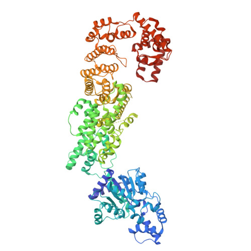
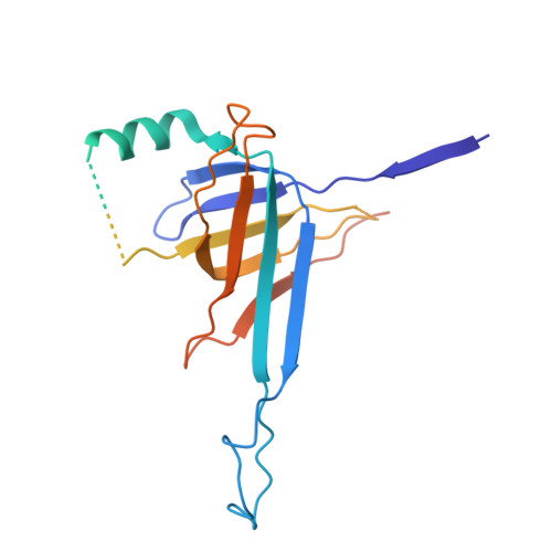
The structural basis of the activation and inhibition of DSR2 NADase by phage proteins.

Wang, R.Xu, Q.Wu, Z.Li, J.Guo, H.Liao, T.Shi, Y.Yuan, L.Gao, H.Yang, R.Shi, Z.Li, F.

(2024) Nat Commun 15: 6185-6185

  • DOI: https://doi.org/10.1038/s41467-024-50410-0
  • Primary Citation of Related Structures:  
    8Y13, 8Y34, 8Y3M, 8Y3W, 8Y3Y, 8ZC9

  • PubMed Abstract: 

    DSR2, a Sir2 domain-containing protein, protects bacteria from phage infection by hydrolyzing NAD + . The enzymatic activity of DSR2 is triggered by the SPR phage tail tube protein (TTP), while suppressed by the SPbeta phage-encoded DSAD1 protein, enabling phages to evade the host defense. However, the molecular mechanisms of activation and inhibition of DSR2 remain elusive. Here, we report the cryo-EM structures of apo DSR2, DSR2-TTP-NAD + and DSR2-DSAD1 complexes. DSR2 assembles into a head-to-head tetramer mediated by its Sir2 domain. The C-terminal helical regions of DSR2 constitute four partner-binding cavities with opened and closed conformation. Two TTP molecules bind to two of the four C-terminal cavities, inducing conformational change of Sir2 domain to activate DSR2. Furthermore, DSAD1 competes with the activator for binding to the C-terminal cavity of DSR2, effectively suppressing its enzymatic activity. Our results provide the mechanistic insights into the DSR2-mediated anti-phage defense system and DSAD1-dependent phage immune evasion.


  • Organizational Affiliation

    MOE Key Laboratory of Rare Pediatric Diseases, Center for Medical Genetics, School of Life Sciences, Central South University, Changsha, Hunan, China.


Macromolecules
Find similar proteins by:  (by identity cutoff)  |  3D Structure
Entity ID: 1
MoleculeChains Sequence LengthOrganismDetailsImage
SIR2-like domain-containing proteinA [auth B],
B [auth A],
D [auth E],
E [auth D]
1,005Bacillus subtilisMutation(s): 0 
Gene Names: BSNT_07056
UniProt
Find proteins for D4G637 (Bacillus subtilis subsp. natto (strain BEST195))
Explore D4G637 
Go to UniProtKB:  D4G637
Entity Groups  
Sequence Clusters30% Identity50% Identity70% Identity90% Identity95% Identity100% Identity
UniProt GroupD4G637
Sequence Annotations
Expand
  • Reference Sequence
Find similar proteins by:  (by identity cutoff)  |  3D Structure
Entity ID: 2
MoleculeChains Sequence LengthOrganismDetailsImage
tail tube protein
C, F
264Bacillus subtilisMutation(s): 0 
Gene Names: J5227_09585
UniProt
Find proteins for A0A162TY69 (Bacillus subtilis)
Explore A0A162TY69 
Go to UniProtKB:  A0A162TY69
Entity Groups  
Sequence Clusters30% Identity50% Identity70% Identity90% Identity95% Identity100% Identity
UniProt GroupA0A162TY69
Sequence Annotations
Expand
  • Reference Sequence
Experimental Data & Validation

Experimental Data

  • Method: ELECTRON MICROSCOPY
  • Resolution: 3.14 Å
  • Aggregation State: PARTICLE 
  • Reconstruction Method: SINGLE PARTICLE 

Structure Validation

View Full Validation Report


Entry History & Funding Information

Deposition Data


Funding OrganizationLocationGrant Number
National Natural Science Foundation of China (NSFC)China32370742
National Natural Science Foundation of China (NSFC)China32271264
National Natural Science Foundation of China (NSFC)China32200129

Revision History  (Full details and data files)

  • Version 1.0: 2024-09-11
    Type: Initial release