


Crystallographic analysis of MitM, which catalyzes the post-mitosane modification in mitomycin biosynthesis
Xia, M., Dong, D.To be published.
Experimental Data Snapshot
Starting Model: experimental
View more details
Entity ID: 1 | |||||
|---|---|---|---|---|---|
| Molecule | Chains | Sequence Length | Organism | Details | Image |
| MitM | 283 | Streptomyces caespitosus | Mutation(s): 0 Gene Names: mitM | 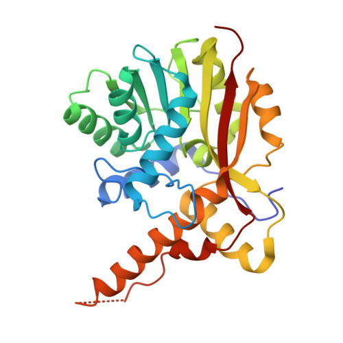 | |
UniProt | |||||
Entity Groups | |||||
| Sequence Clusters | 30% Identity50% Identity70% Identity90% Identity95% Identity100% Identity | ||||
| UniProt Group | Q9X5Q9 | ||||
Sequence AnnotationsExpand | |||||
Reference Sequence | |||||
| Ligands 2 Unique | |||||
|---|---|---|---|---|---|
| ID | Chains | Name / Formula / InChI Key | 2D Diagram | 3D Interactions | |
| SAH (Subject of Investigation/LOI) Download:Ideal Coordinates CCD File | C [auth A], D [auth B] | S-ADENOSYL-L-HOMOCYSTEINE C14 H20 N6 O5 S ZJUKTBDSGOFHSH-WFMPWKQPSA-N |  | ||
| MQA (Subject of Investigation/LOI) Download:Ideal Coordinates CCD File | E [auth B] | [(1aS,8S,8aR,8bS)-6,8a-dimethoxy-5-methyl-4,7-dioxo-1,1a,2,4,7,8,8a,8b-octahydroazireno[2',3':3,4]pyrrolo[1,2-a]indol-8-yl]methyl carbamate C16 H19 N3 O6 HYFMSAFINFJTFH-NGSRAFSJSA-N |  | ||
| Length ( Å ) | Angle ( ˚ ) |
|---|---|
| a = 113.953 | α = 90 |
| b = 113.953 | β = 90 |
| c = 108.771 | γ = 120 |
| Software Name | Purpose |
|---|---|
| PHENIX | refinement |
| SCALA | data scaling |
| PHASER | phasing |
| PDB_EXTRACT | data extraction |
| MOSFLM | data reduction |
| Funding Organization | Location | Grant Number |
|---|---|---|
| Ministry of Science and Technology (MoST, China) | China | 2022YFC2303100 |


