8Z8B | pdb_00008z8b

Cryo-EM structure of human HCAR1-Gi complex without ligand (apo state)


Experimental Data Snapshot

  • Method: ELECTRON MICROSCOPY
  • Resolution: 2.65 Å
  • Aggregation State: PARTICLE 
  • Reconstruction Method: SINGLE PARTICLE 

wwPDB Validation 3D Report Full Report

Validation slider image for 8Z8B

This is version 1.1 of the entry. See complete history

Literature

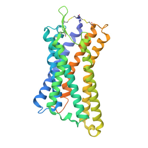
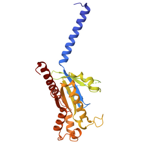
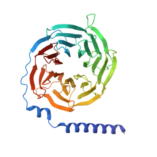
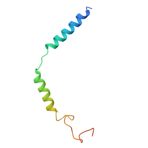
Structural insights into the activation mechanism of the human metabolite receptor HCAR1.

Gao, M.Zang, S.Zhu, Y.Xi, K.Du, Y.Cheng, S.Miao, L.Lu, Y.Mao, C.Zhang, Y.Ma, X.

(2026) Sci Signal 19: eadw1483-eadw1483

  • DOI: https://doi.org/10.1126/scisignal.adw1483
  • Primary Citation Related Structures: 
    8Z87, 8Z8A, 8Z8B

  • PubMed Abstract: 

    Hydroxycarboxylic acid receptor 1 (HCAR1) is a class A G protein-coupled receptor (GPCR) that is activated by the endogenous metabolite l-lactate and that plays an important role in various metabolic and inflammatory disorders. HCAR1 uses distinct ligand recognition and self-activation mechanisms to mediate specific pathophysiological functions through Gα i/o and β-arrestin signaling pathways. To support effective drug development targeting HCAR1, we investigated ligand recognition and activation mechanisms through cryo-electron microscopy (cryo-EM) structures of the HCAR1-Gα i1 complex in the apo state or with l-lactate or with the synthetic agonist CHBA. Compared with other HCARs, HCAR1 has a more compact binding pocket, which is stabilized by three unique disulfide bonds. l-lactate exhibited a flexible binding mode and relatively weak intermolecular interactions, thus requiring millimolar concentrations for receptor activation. In contrast, the binding of CHBA was more stable because of its chlorinated benzene ring, thus resulting in improved agonist potency. Structural comparisons with HCAR2 identified critical residues that restrict the size of the binding pocket of HCAR1 and influence ligand selectivity. Self-activation of HCAR1 is driven by conformational rearrangements within extracellular loop 2, with Phe168 ECL2 playing a pivotal role as the key agonist. Together, these results clarify the mechanisms underlying HCAR1 activation, self-activation, and ligand selectivity, providing a structural framework for the design of high-affinity, selective agonists and inverse agonists with minimized off-target effects.


  • Organizational Affiliation
    • MOE Medical Basic Research Innovation Center for Gut Microbiota and Chronic Diseases, School of Medicine, Jiangnan University, Wuxi 214122, China.

Macromolecule Content 

  • Total Structure Weight: 152.01 kDa 
  • Atom Count: 8,875 
  • Modeled Residue Count: 1,137 
  • Deposited Residue Count: 1,365 
  • Unique protein chains: 5

Macromolecules

Find similar proteins by:|  3D Structure
Entity ID: 1
MoleculeChains  Sequence LengthOrganismDetailsImage
Hydroxycarboxylic acid receptor 1A [auth R]346Homo sapiensMutation(s): 0 
Gene Names: HCAR1GPR104GPR81HCA1FKSG80
UniProt & NIH Common Fund Data Resources
Find proteins for Q9BXC0 (Homo sapiens)
Explore Q9BXC0 
Go to UniProtKB:  Q9BXC0
PHAROS:  Q9BXC0
GTEx:  ENSG00000196917 
Entity Groups
Sequence Clusters30% Identity50% Identity70% Identity90% Identity95% Identity100% Identity
UniProt GroupQ9BXC0
Sequence Annotations
Expand
Reference Sequence
Find similar proteins by:|  3D Structure
Entity ID: 2
MoleculeChains  Sequence LengthOrganismDetailsImage
Guanine nucleotide-binding protein G(i) subunit alpha-1B [auth A]354Homo sapiensMutation(s): 0 
Gene Names: GNAI1
EC: 3.6.5
UniProt & NIH Common Fund Data Resources
Find proteins for P63096 (Homo sapiens)
Explore P63096 
Go to UniProtKB:  P63096
PHAROS:  P63096
GTEx:  ENSG00000127955 
Entity Groups
Sequence Clusters30% Identity50% Identity70% Identity90% Identity95% Identity100% Identity
UniProt GroupP63096
Sequence Annotations
Expand
Reference Sequence
Find similar proteins by:|  3D Structure
Entity ID: 3
MoleculeChains  Sequence LengthOrganismDetailsImage
Guanine nucleotide-binding protein G(I)/G(S)/G(T) subunit beta-1C [auth B]345Homo sapiensMutation(s): 0 
Gene Names: GNB1
UniProt & NIH Common Fund Data Resources
Find proteins for P62873 (Homo sapiens)
Explore P62873 
Go to UniProtKB:  P62873
PHAROS:  P62873
GTEx:  ENSG00000078369 
Entity Groups
Sequence Clusters30% Identity50% Identity70% Identity90% Identity95% Identity100% Identity
UniProt GroupP62873
Sequence Annotations
Expand
Reference Sequence
Find similar proteins by:|  3D Structure
Entity ID: 4
MoleculeChains  Sequence LengthOrganismDetailsImage
Guanine nucleotide-binding protein G(I)/G(S)/G(O) subunit gamma-2D [auth G]70Homo sapiensMutation(s): 0 
Gene Names: GNG2
UniProt & NIH Common Fund Data Resources
Find proteins for P59768 (Homo sapiens)
Explore P59768 
Go to UniProtKB:  P59768
PHAROS:  P59768
GTEx:  ENSG00000186469 
Entity Groups
Sequence Clusters30% Identity50% Identity70% Identity90% Identity95% Identity100% Identity
UniProt GroupP59768
Sequence Annotations
Expand
Reference Sequence
Find similar proteins by:|  3D Structure
Entity ID: 5
MoleculeChains  Sequence LengthOrganismDetailsImage
single Fab chain (scFv16)E [auth S]250synthetic constructMutation(s): 0 

Experimental Data & Validation

Experimental Data

  • Method: ELECTRON MICROSCOPY
  • Resolution: 2.65 Å
  • Aggregation State: PARTICLE 
  • Reconstruction Method: SINGLE PARTICLE 

Structure Validation

View Full Validation Report


Entry History 

& Funding Information

Deposition Data


Funding OrganizationLocationGrant Number
Not funded--

Revision History  (Full details and data files)

  • Version 1.0: 2025-10-29
    Type: Initial release
  • Version 1.1: 2026-05-13
    Changes: Data collection, Database references