8Z7M | pdb_00008z7m

The crystal structure of AFM-1


Experimental Data Snapshot

  • Method: X-RAY DIFFRACTION
  • Resolution: 2.60 Å
  • R-Value Free: 
    0.258 (Depositor), 0.252 (DCC) 
  • R-Value Work: 
    0.210 (Depositor), 0.216 (DCC) 
  • R-Value Observed: 
    0.214 (Depositor) 

wwPDB Validation 3D Report Full Report

Validation slider image for 8Z7M

This is version 1.0 of the entry. See complete history

Literature

Structural insight into the subclass B1 metallo-beta-lactamase AFM-1.

Niu, W.Ti, R.Li, D.Dong, R.Dong, J.Ye, Y.Xiao, Y.Wang, Z.

(2024) Biochem Biophys Res Commun 720: 150102-150102

  • DOI: https://doi.org/10.1016/j.bbrc.2024.150102
  • Primary Citation Related Structures: 
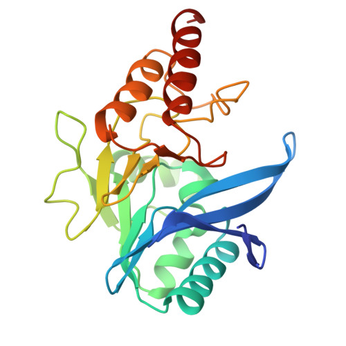
    8Z7M

  • PubMed Abstract: 

    The emergence of drug-resistant bacteria, facilitated by metallo-beta-lactamases (MBLs), presents a significant obstacle to the effective use of antibiotics in the management of clinical drug-resistant bacterial infections. AFM-1 is a MBL derived from Alcaligenes faecalis and shares 86% homology with the NDM-1 family. Both AFM-1 and NDM-1 demonstrate the ability to hydrolyze ampicillin and other β-lactam antibiotics, however, their substrate affinities vary, and the specific reason for this variation remains unknown. We present the high-resolution structure of AFM-1. The active center of AFM-1 binds two zinc ions, and the conformation of the key amino acid residues in the active center is in accordance with that of NDM-1. However, the substrate-binding pocket of AFM-1 is considerably smaller than that of NDM-1. Additionally, the mutation of amino acid residues in the Loop3 region, as compared to NDM-1, results in the formation of a dense hydrophobic patch comprised of hydrophobic amino acid residues in this area, which facilitates substrate binding. Our findings lay the foundation for understanding the molecular mechanism of AFM-1 with a high affinity for substrates and provide a novel theoretical foundation for addressing the issue of drug resistance caused by B1 MBLs.


  • Organizational Affiliation
    • School of Life Sciences, Tianjin University, Tianjin, 300072, China.

Macromolecule Content 

  • Total Structure Weight: 51.37 kDa 
  • Atom Count: 3,569 
  • Modeled Residue Count: 462 
  • Deposited Residue Count: 480 
  • Unique protein chains: 1

Macromolecules

Find similar proteins by:|  3D Structure
Entity ID: 1
MoleculeChains  Sequence LengthOrganismDetailsImage
Metallo-beta-lactamase type 2
A, B
240Alcaligenes faecalisMutation(s): 0 
Gene Names: blaAFM
EC: 3.5.2.6
UniProt
Find proteins for A0A3G5BD68 (Alcaligenes faecalis)
Explore A0A3G5BD68 
Go to UniProtKB:  A0A3G5BD68
Entity Groups
Sequence Clusters30% Identity50% Identity70% Identity90% Identity95% Identity100% Identity
UniProt GroupA0A3G5BD68
Sequence Annotations
Expand
Reference Sequence

Experimental Data & Validation

Experimental Data

  • Method: X-RAY DIFFRACTION
  • Resolution: 2.60 Å
  • R-Value Free:  0.258 (Depositor), 0.252 (DCC) 
  • R-Value Work:  0.210 (Depositor), 0.216 (DCC) 
  • R-Value Observed: 0.214 (Depositor) 
Space Group: P 21 21 21
Unit Cell:
Length ( Å )Angle ( ˚ )
a = 48.96α = 90
b = 105.459β = 90
c = 106.359γ = 90
Software Package:
Software NamePurpose
PHENIXrefinement
XDSdata scaling
PHENIXmodel building
PDB_EXTRACTdata extraction

Structure Validation

View Full Validation Report


Entry History 

& Funding Information

Deposition Data


Funding OrganizationLocationGrant Number
National Natural Science Foundation of China (NSFC)China82170680

Revision History  (Full details and data files)

  • Version 1.0: 2025-02-26
    Type: Initial release