


Crystal Structure of Protein Glutaminase FBPG(Flavobacterium sp.316)
Long, Y.T., Lan, D.M., Wang, Y.H.To be published.
Experimental Data Snapshot
Starting Model: in silico
View more details
wwPDB Validation 3D Report Full Report

Entity ID: 1 | |||||
|---|---|---|---|---|---|
| Molecule | Chains | Sequence Length | Organism | Details | Image |
| Protein glutaminase domain-containing protein | 312 | Flavobacterium sp. 316 | Mutation(s): 0 Gene Names: SY27_02215 | 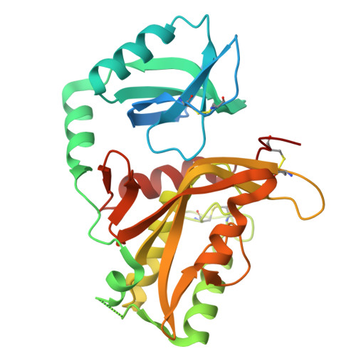 | |
| Length ( Å ) | Angle ( ˚ ) |
|---|---|
| a = 51.338 | α = 90 |
| b = 52.455 | β = 90 |
| c = 109.467 | γ = 90 |
| Software Name | Purpose |
|---|---|
| PHENIX | refinement |
| HKL-3000 | data reduction |
| HKL-3000 | data scaling |
| PHENIX | phasing |
| Funding Organization | Location | Grant Number |
|---|---|---|
| Not funded | -- |


