8Z1Y | pdb_00008z1y

Cryo-EM structure of Escherichia coli DppABCDF in the pre-catalytic state


Experimental Data Snapshot

  • Method: ELECTRON MICROSCOPY
  • Resolution: 2.73 Å
  • Aggregation State: PARTICLE 
  • Reconstruction Method: SINGLE PARTICLE 

wwPDB Validation   3D Report Full Report


This is version 1.1 of the entry. See complete history


Literature

Structural characterization of the ABC transporter DppABCDF in Escherichia coli reveals insights into dipeptide acquisition.

Li, P.Zhang, M.Huang, Y.

(2025) PLoS Biol 23: e3003026-e3003026

  • DOI: https://doi.org/10.1371/journal.pbio.3003026
  • Primary Citation of Related Structures:  
    8Z1V, 8Z1W, 8Z1X, 8Z1Y

  • PubMed Abstract: 

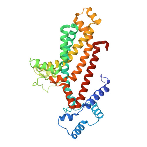
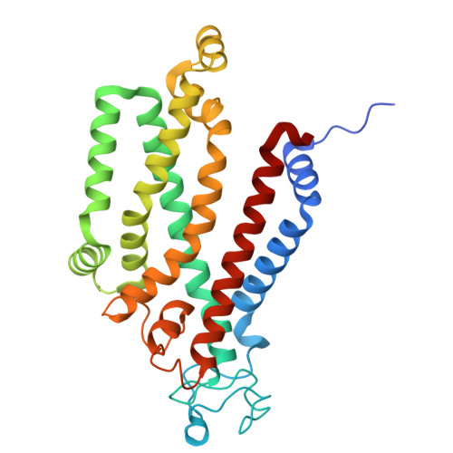
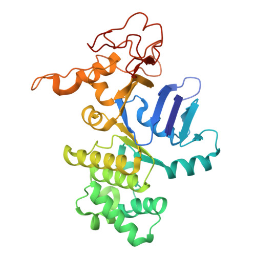
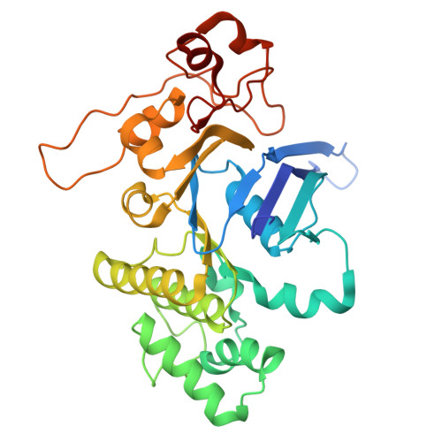
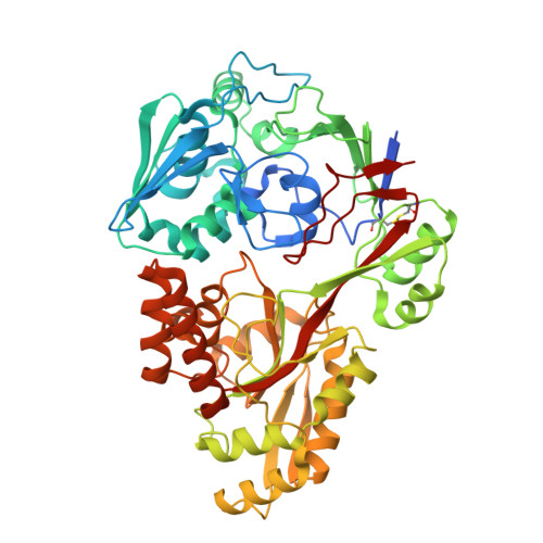
    The prokaryote-specific ATP-binding cassette (ABC) peptide transporters are involved in various physiological processes and plays an important role in transporting naturally occurring antibiotics across the membrane to their intracellular targets. The dipeptide transporter DppABCDF in Gram-negative bacteria is composed of five distinct subunits, yet its assembly and underlying peptide import mechanism remain elusive. Here, we report the cryo-EM structures of the DppBCDF translocator from Escherichia coli in both its apo form and in complexes bound to nonhydrolyzable or slowly hydrolyzable ATP analogs (AMPPNP and ATPγS), as well as the ATPγS-bound DppABCDF full transporter. Unlike the reported heterotrimeric Mycobacterium tuberculosis DppBCD translocator, the E. coli DppBCDF translocator is a heterotetramer, with a [4Fe-4S] cluster at the C-terminus of each ATPase subunit. Structural studies reveal that ATPγS/AMPPNP-bound DppBCDF adopts an inward-facing conformation, similar to that of apo-DppBCDF, with only one ATPγS or AMPPNP molecule bound to DppF. By contrast, ATPγS-bound DppABCDF adopts an outward-facing conformation, with two ATPγS molecules glueing DppD and DppF at the interface. Consistent with structural observations, ATPase activity assays show that the DppBCDF translocator itself is inactive and its activation requires concurrent binding of DppA and ATP. In addition, bacterial complementation experiments imply that a unique periplasmic scoop motif in DppB may play important roles in ensuring dipeptide substrates import across the membrane, presumably by preventing dipeptide back-and-forth binding to DppA and avoiding dipeptides escaping into the periplasm upon being released from DppA.


  • Organizational Affiliation

    National Laboratory of Biomacromolecules, CAS Center for Excellence in Biomacromolecules, Institute of Biophysics, Chinese Academy of Sciences, Beijing, China.


Macromolecules
Find similar proteins by:  (by identity cutoff)  |  3D Structure
Entity ID: 1
MoleculeChains Sequence LengthOrganismDetailsImage
Dipeptide transport system permease protein DppB339Escherichia coli K-12Mutation(s): 0 
Gene Names: dppBb3543JW3512
Membrane Entity: Yes 
UniProt
Find proteins for P0AEF8 (Escherichia coli (strain K12))
Explore P0AEF8 
Go to UniProtKB:  P0AEF8
Entity Groups  
Sequence Clusters30% Identity50% Identity70% Identity90% Identity95% Identity100% Identity
UniProt GroupP0AEF8
Sequence Annotations
Expand
  • Reference Sequence
Find similar proteins by:  (by identity cutoff)  |  3D Structure
Entity ID: 2
MoleculeChains Sequence LengthOrganismDetailsImage
Dipeptide transport system permease protein DppC300Escherichia coli K-12Mutation(s): 0 
Gene Names: dppCb3542JW3511
Membrane Entity: Yes 
UniProt
Find proteins for P0AEG1 (Escherichia coli (strain K12))
Explore P0AEG1 
Go to UniProtKB:  P0AEG1
Entity Groups  
Sequence Clusters30% Identity50% Identity70% Identity90% Identity95% Identity100% Identity
UniProt GroupP0AEG1
Sequence Annotations
Expand
  • Reference Sequence
Find similar proteins by:  (by identity cutoff)  |  3D Structure
Entity ID: 3
MoleculeChains Sequence LengthOrganismDetailsImage
Dipeptide transport ATP-binding protein DppD327Escherichia coli K-12Mutation(s): 0 
Gene Names: dppDb3541JW3510
EC: 7.4.2.9
Membrane Entity: Yes 
UniProt
Find proteins for P0AAG0 (Escherichia coli (strain K12))
Explore P0AAG0 
Go to UniProtKB:  P0AAG0
Entity Groups  
Sequence Clusters30% Identity50% Identity70% Identity90% Identity95% Identity100% Identity
UniProt GroupP0AAG0
Sequence Annotations
Expand
  • Reference Sequence
Find similar proteins by:  (by identity cutoff)  |  3D Structure
Entity ID: 4
MoleculeChains Sequence LengthOrganismDetailsImage
Dipeptide transport ATP-binding protein DppF334Escherichia coli K-12Mutation(s): 0 
Gene Names: dppFdppEb3540JW3509
EC: 7.4.2.9
Membrane Entity: Yes 
UniProt
Find proteins for P37313 (Escherichia coli (strain K12))
Explore P37313 
Go to UniProtKB:  P37313
Entity Groups  
Sequence Clusters30% Identity50% Identity70% Identity90% Identity95% Identity100% Identity
UniProt GroupP37313
Sequence Annotations
Expand
  • Reference Sequence
Find similar proteins by:  (by identity cutoff)  |  3D Structure
Entity ID: 5
MoleculeChains Sequence LengthOrganismDetailsImage
Dipeptide-binding proteinE [auth F]535Escherichia coli K-12Mutation(s): 0 
Gene Names: dppAb3544JW3513
Membrane Entity: Yes 
UniProt
Find proteins for P23847 (Escherichia coli (strain K12))
Explore P23847 
Go to UniProtKB:  P23847
Entity Groups  
Sequence Clusters30% Identity50% Identity70% Identity90% Identity95% Identity100% Identity
UniProt GroupP23847
Sequence Annotations
Expand
  • Reference Sequence
Experimental Data & Validation

Experimental Data

  • Method: ELECTRON MICROSCOPY
  • Resolution: 2.73 Å
  • Aggregation State: PARTICLE 
  • Reconstruction Method: SINGLE PARTICLE 

Structure Validation

View Full Validation Report


Entry History & Funding Information

Deposition Data


Funding OrganizationLocationGrant Number
Chinese Academy of SciencesChina--

Revision History  (Full details and data files)

  • Version 1.0: 2025-01-29
    Type: Initial release
  • Version 1.1: 2025-03-19
    Changes: Data collection, Database references