8Z18

The tetramer complex of DSR2 and tube-forming domain of phage tail tube protein


Experimental Data Snapshot

  • Method: ELECTRON MICROSCOPY
  • Resolution: 3.94 Å
  • Aggregation State: PARTICLE 
  • Reconstruction Method: SINGLE PARTICLE 

wwPDB Validation   3D Report Full Report


This is version 1.0 of the entry. See complete history


Literature

Structural insights into autoinhibition and activation of defense-associated sirtuin protein.

Yang, X.Wang, Y.Zheng, J.

(2024) Int J Biol Macromol 277: 134145-134145

  • DOI: https://doi.org/10.1016/j.ijbiomac.2024.134145
  • Primary Citation of Related Structures:  
    8YKF, 8YL5, 8YLN, 8YLT, 8Z18, 8ZTR

  • PubMed Abstract: 

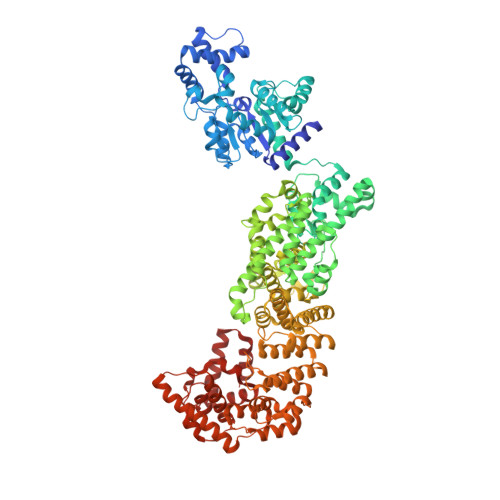
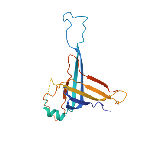
    Bacterial defense-associated sirtuin 2 (DSR2) proteins harbor an N-terminal sirtuin (SIR2) domain degrading NAD + . DSR2 from Bacillus subtilis 29R is autoinhibited and unable to hydrolyze NAD + in the absence of phage infection. A tail tube protein (TTP) of phage SPR activates the DSR2 while a DSR2-inhibiting protein of phage SPbeta, known as DSAD1 (DSR anti-defense 1), inactivates the DSR2. Although DSR2 structures in complexed with TTP and DSAD1, respectively, have been reported recently, the autoinhibition and activation mechanisms remain incompletely understood. Here, we present cryo-electron microscopy structures of the DSR2-NAD + complex in autoinhibited state and the in vitro assembled DSR2-TFD (TTP tube-forming domain) complex in activated state. The DSR2-NAD + complex reveals that the autoinhibited DSR2 assembles into an inactive tetramer, binding NAD + through a distinct pocket situated outside active site. Binding of TFD into cavities within the sensor domains of DSR2 triggers a conformational change in SIR2 regions, activating its NADase activity, whereas the TTP β-sandwich domain (BSD) is flexible and does not contribute to the activation process. The activated form of DSR2 exists as tetramers and dimers, with the tetramers exhibiting more NADase activity. Overall, our results extend the current understanding of autoinhibition and activation of DSR2 immune proteins.


  • Organizational Affiliation

    State Key Laboratory of Microbial Metabolism, School of Life Sciences and Biotechnology, Shanghai Jiao Tong University, Shanghai 200240, China.


Macromolecules
Find similar proteins by:  (by identity cutoff)  |  3D Structure
Entity ID: 1
MoleculeChains Sequence LengthOrganismDetailsImage
SIR2-like domain-containing protein
A, B, C, D
1,005Bacillus subtilis subsp. natto BEST195Mutation(s): 0 
Gene Names: BSNT_07056
UniProt
Find proteins for D4G637 (Bacillus subtilis subsp. natto (strain BEST195))
Explore D4G637 
Go to UniProtKB:  D4G637
Entity Groups  
Sequence Clusters30% Identity50% Identity70% Identity90% Identity95% Identity100% Identity
UniProt GroupD4G637
Sequence Annotations
Expand
  • Reference Sequence
Find similar proteins by:  (by identity cutoff)  |  3D Structure
Entity ID: 2
MoleculeChains Sequence LengthOrganismDetailsImage
Bacillus phage SPR Tube protein
E, F, G, H
264Bacillus phage SPRMutation(s): 0 
Gene Names: B4122_1986
UniProt
Find proteins for A0A162TY69 (Bacillus subtilis)
Explore A0A162TY69 
Go to UniProtKB:  A0A162TY69
Entity Groups  
Sequence Clusters30% Identity50% Identity70% Identity90% Identity95% Identity100% Identity
UniProt GroupA0A162TY69
Sequence Annotations
Expand
  • Reference Sequence
Experimental Data & Validation

Experimental Data

  • Method: ELECTRON MICROSCOPY
  • Resolution: 3.94 Å
  • Aggregation State: PARTICLE 
  • Reconstruction Method: SINGLE PARTICLE 
EM Software:
TaskSoftware PackageVersion
MODEL REFINEMENTPHENIX1.20.1_4487:

Structure Validation

View Full Validation Report


Entry History & Funding Information

Deposition Data


Funding OrganizationLocationGrant Number
National Natural Science Foundation of China (NSFC)China32070040, 32370071

Revision History  (Full details and data files)

  • Version 1.0: 2024-08-14
    Type: Initial release