8YX7 | pdb_00008yx7

Structure of a Cys-loop Receptor under Acidic Condition


Experimental Data Snapshot

  • Method: ELECTRON MICROSCOPY
  • Resolution: 2.80 Å
  • Aggregation State: PARTICLE 
  • Reconstruction Method: SINGLE PARTICLE 

wwPDB Validation   3D Report Full Report


This is version 1.2 of the entry. See complete history


Literature

Structural insights into the activation mechanism of the human zinc-activated channel.

Lu, X.Li, D.Wang, Y.Zhang, G.Wen, T.Lu, Y.Jia, N.Wang, X.Chang, S.Zhang, X.Lin, J.Chen, Y.H.Yang, X.Shen, Y.

(2025) Nat Commun 16: 442-442

  • DOI: https://doi.org/10.1038/s41467-024-55807-5
  • Primary Citation of Related Structures:  
    8YX6, 8YX7, 8YX8

  • PubMed Abstract: 

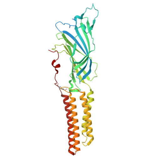
    The zinc-activated channel (ZAC) is an atypical mammalian cys-loop receptor (CLR) that is activated by zinc ions and protons, allowing cations to pass through. The molecular mechanism that ligands use to activate ZAC remains elusive. Here, we present three cryo-electron microscopy reconstructions of human ZAC (hZAC) under different conditions. These three hZAC structures display highly similar conformations to one another, forming symmetrical homo-pentamers with a central ion-conduction pore. The hZAC protomer comprises an extracellular domain (ECD) and a transmembrane domain (TMD), sharing more structural similarity with anion-permeable CLRs, such as glycine receptors and type A γ-aminobutyric acid receptors. Notably, hZAC possesses a distinctive C-tail that establishes a disulfide bond with the loop M2-M3 in the TMD and occupies what is typically the canonical neurotransmitter orthosteric site in other mammalian CLRs. Moreover, the tip of the cys-loop creates an unprecedented orthosteric site in hZAC. The binding of Zn 2+ triggers a conformational shift in the cys-loop, which presumably prompts the loop M2-M3 to move and open the channel gate. This study sheds light on the assembly of the channel, its structural features, and the process of signal transduction in hZAC.


  • Organizational Affiliation
    • State Key Laboratory of Medicinal Chemical Biology and Frontiers Science Center for Cell Responses, College of Life Sciences, Nankai University, Tianjin, 300350, China.

Macromolecules
Find similar proteins by:  (by identity cutoff)  |  3D Structure
Entity ID: 1
MoleculeChains Sequence LengthOrganismDetailsImage
Ligand-gated cation channel ZACN
A, B, C, D, E
420Homo sapiensMutation(s): 0 
Gene Names: ZACNL2LGICZLGICZ1ZAC
Membrane Entity: Yes 
UniProt & NIH Common Fund Data Resources
Find proteins for Q401N2 (Homo sapiens)
Explore Q401N2 
Go to UniProtKB:  Q401N2
PHAROS:  Q401N2
GTEx:  ENSG00000186919 
Entity Groups  
Sequence Clusters30% Identity50% Identity70% Identity90% Identity95% Identity100% Identity
UniProt GroupQ401N2
Glycosylation
Glycosylation Sites: 2Go to GlyGen: Q401N2-1
Sequence Annotations
Expand
  • Reference Sequence
Oligosaccharides

Help

Entity ID: 2
MoleculeChains Length2D Diagram Glycosylation3D Interactions
2-acetamido-2-deoxy-beta-D-glucopyranose-(1-4)-2-acetamido-2-deoxy-beta-D-glucopyranose
F, G, H, I, J
2N-Glycosylation
Glycosylation Resources
GlyTouCan:  G42666HT
GlyCosmos:  G42666HT
GlyGen:  G42666HT
Experimental Data & Validation

Experimental Data

  • Method: ELECTRON MICROSCOPY
  • Resolution: 2.80 Å
  • Aggregation State: PARTICLE 
  • Reconstruction Method: SINGLE PARTICLE 
EM Software:
TaskSoftware PackageVersion
MODEL REFINEMENTPHENIX

Structure Validation

View Full Validation Report


Entry History & Funding Information

Deposition Data


Funding OrganizationLocationGrant Number
National Natural Science Foundation of China (NSFC)China--

Revision History  (Full details and data files)

  • Version 1.0: 2025-01-01
    Type: Initial release
  • Version 1.1: 2025-07-16
    Changes: Data collection, Database references
  • Version 1.2: 2025-07-23
    Changes: Data collection