8YRT | pdb_00008yrt

Crystal structure of D-amino acid transaminase from Haliscomenobacter hydrossis in the holo form obtained at pH 7.0


Experimental Data Snapshot

  • Method: X-RAY DIFFRACTION
  • Resolution: 2.00 Å
  • R-Value Free: 
    0.190 (Depositor), 0.201 (DCC) 
  • R-Value Work: 
    0.145 (Depositor), 0.158 (DCC) 
  • R-Value Observed: 
    0.147 (Depositor) 

Starting Model: experimental
View more details

wwPDB Validation 3D Report Full Report

Validation slider image for 8YRT

Ligand Structure Quality Assessment 


This is version 1.1 of the entry. See complete history

Literature

Incorporation of pyridoxal-5'-phosphate into the apoenzyme: A structural study of D-amino acid transaminase from Haliscomenobacter hydrossis.

Bakunova, A.K.Matyuta, I.O.Minyaev, M.E.Boyko, K.M.Popov, V.O.Bezsudnova, E.Y.

(2024) Biochim Biophys Acta Proteins Proteom 1873: 141056-141056

  • DOI: https://doi.org/10.1016/j.bbapap.2024.141056
  • Primary Citation Related Structures: 
    7P8O, 8YRT, 8YRU

  • PubMed Abstract: 

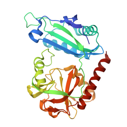
    Pyridoxal-5'-phosphate (PLP)-dependent transaminases are key enzymes of amino acid metabolism in cells and remarkable biocatalysts of stereoselective amination for process chemistry applications. As cofactor-dependent enzymes, transaminases are prone to cofactor leakage. Here we discuss the holoenzyme-apoenzyme interconversion and the kinetics of PLP incorporation into the apo form of a PLP-dependent transaminase from Haliscomenobacter hydrossis. PLP binding to the apoenzyme was slow in buffer, but was accelerated in the presence of substrates. Two crystal structures of the apoenzyme were obtained: the directly obtained apoenzyme (PDB ID: 7P8O) and the one obtained by soaking crystals of the holoenzyme in a phenylhydrazine solution (PDB ID: 8YRU). The mechanism of PLP association with the apoenzyme was proposed on the basis of structural analysis of these apo forms. Three rearrangement steps, including (I) anchoring of the PLP via the phosphate group, (II) displacement of two loops, and (III) Schiff-bonding between the PLP and the ε-amino group of the catalytic lysine residue, reconstituted the active holo form of the transaminase from H. hydrossis. The results obtained allowed us to determine in the active site a permanent part and elements that are assembled by PLP, these findings may be useful for transaminase engineering for biocatalysis.


  • Organizational Affiliation
    • Bach Institute of Biochemistry, Research Centre of Biotechnology of the Russian Academy of Sciences, Moscow, Russia.

Macromolecule Content 

  • Total Structure Weight: 32.57 kDa 
  • Atom Count: 2,446 
  • Modeled Residue Count: 282 
  • Deposited Residue Count: 283 
  • Unique protein chains: 1

Macromolecules

Find similar proteins by:|  3D Structure
Entity ID: 1
MoleculeChains  Sequence LengthOrganismDetailsImage
Aminotransferase class IV283Haliscomenobacter hydrossis DSM 1100Mutation(s): 0 
Gene Names: Halhy_2446
EC: 2.6.1.42
UniProt
Find proteins for F4KWH0 (Haliscomenobacter hydrossis (strain ATCC 27775 / DSM 1100 / LMG 10767 / O))
Explore F4KWH0 
Go to UniProtKB:  F4KWH0
Entity Groups
Sequence Clusters30% Identity50% Identity70% Identity90% Identity95% Identity100% Identity
UniProt GroupF4KWH0
Sequence Annotations
Expand
Reference Sequence

Small Molecules

Ligands 1 Unique
IDChains Name / Formula / InChI Key2D Diagram3D Interactions
PLP
(Subject of Investigation/LOI)

Query on PLP


Download:Ideal Coordinates CCD File
B [auth A]PYRIDOXAL-5'-PHOSPHATE
C8 H10 N O6 P
NGVDGCNFYWLIFO-UHFFFAOYSA-N

Experimental Data & Validation

Experimental Data

  • Method: X-RAY DIFFRACTION
  • Resolution: 2.00 Å
  • R-Value Free:  0.190 (Depositor), 0.201 (DCC) 
  • R-Value Work:  0.145 (Depositor), 0.158 (DCC) 
  • R-Value Observed: 0.147 (Depositor) 
Space Group: C 1 2 1
Unit Cell:
Length ( Å )Angle ( ˚ )
a = 86.043α = 90
b = 71.902β = 100.14
c = 52.015γ = 90
Software Package:
Software NamePurpose
REFMACrefinement
Aimlessdata scaling
CrysalisProdata reduction
MOLREPphasing
PDB_EXTRACTdata extraction

Structure Validation

View Full Validation Report


Ligand Structure Quality Assessment 


Entry History 

& Funding Information

Deposition Data


Funding OrganizationLocationGrant Number
Not funded--

Revision History  (Full details and data files)

  • Version 1.0: 2024-04-17
    Type: Initial release
  • Version 1.1: 2024-10-30
    Changes: Advisory, Database references, Derived calculations, Structure summary