8YLU | pdb_00008ylu

structure of phage T6 topoisomerase II central domain bound with DNA


Experimental Data Snapshot

  • Method: ELECTRON MICROSCOPY
  • Resolution: 2.80 Å
  • Aggregation State: PARTICLE 
  • Reconstruction Method: SINGLE PARTICLE 

wwPDB Validation 3D Report Full Report

Validation slider image for 8YLU

This is version 1.1 of the entry. See complete history

Literature

Structural and functional insights into the T-even type bacteriophage topoisomerase II.

Xin, Y.Xian, R.Yang, Y.Cong, J.Rao, Z.Li, X.Chen, Y.

(2024) Nat Commun 15: 8719-8719

  • DOI: https://doi.org/10.1038/s41467-024-53037-3
  • Primary Citation Related Structures: 
    8YLU, 8YO1, 8YO3, 8YO4, 8YO5, 8YO7, 8YO9, 8YOD, 8YON, 9IMJ

  • PubMed Abstract: 

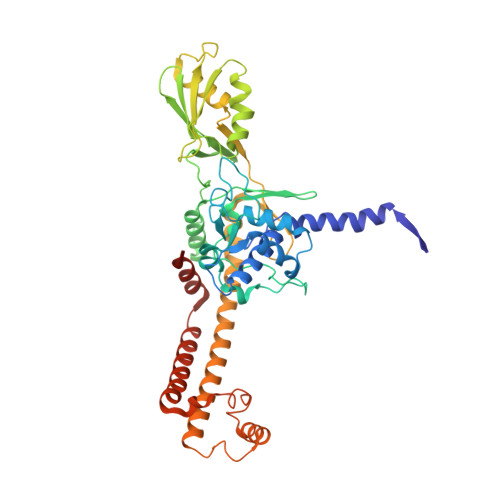
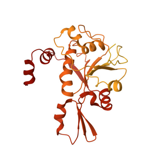
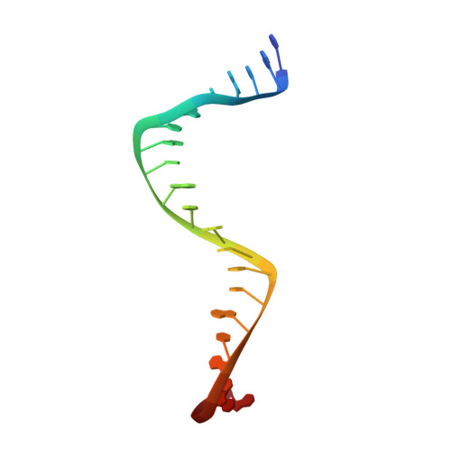
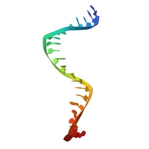
    T-even type bacteriophages are virulent phages commonly used as model organisms, playing a crucial role in understanding various biological processes. One such process involves the regulation of DNA topology during phage replication upon host infection, governed by type IIA DNA topoisomerases. In spite of various studies on prokaryotic and eukaryotic counterparts, viral topoisomerase II remains insufficiently understood, especially the unique domain composition of T4 phage. In this study, we determine the cryo-EM structures of topoisomerase II from T4 and T6 phages, including full-length structures of both apo and DNA-binding states which have never been determined before. Together with other conformational states, these structures provide an explicit blueprint of mechanisms of phage topoisomerase II. Particularly, the asymmetric dimeric interactions observed in cryo-EM structures of T6 phage topoisomerase II ATPase domain and central domain bound with DNA shed light on the asynchronous ATP usage and asynchronous cleavage of the G-segment DNA, respectively. The elucidation of phage topoisomerase II's structures and functions not only enhances our understanding of mechanisms and evolutionary parallels with prokaryotic and eukaryotic homologs but also highlights its potential as a model for developing type IIA topoisomerase inhibitors.


  • Organizational Affiliation
    • National Laboratory of Biomacromolecules, Institute of Biophysics, Chinese Academy of Sciences, Beijing, China.

Macromolecule Content 

  • Total Structure Weight: 255.9 kDa 
  • Atom Count: 11,441 
  • Modeled Residue Count: 1,352 
  • Deposited Residue Count: 2,170 
  • Unique protein chains: 2
  • Unique nucleic acid chains: 2

Macromolecules


Find similar proteins by:|  3D Structure
Entity ID: 1
MoleculeChains  Sequence LengthOrganismDetailsImage
DNA topoisomerase medium subunit
A, B
452Escherichia phage T4Mutation(s): 0 
Gene Names: 52
EC: 5.6.2.2
UniProt
Find proteins for P07065 (Enterobacteria phage T4)
Explore P07065 
Go to UniProtKB:  P07065
Entity Groups
Sequence Clusters30% Identity50% Identity70% Identity90% Identity95% Identity100% Identity
UniProt GroupP07065
Sequence Annotations
Expand
Reference Sequence
Find similar proteins by:|  3D Structure
Entity ID: 2
MoleculeChains  Sequence LengthOrganismDetailsImage
DNA topoisomerase (ATP-hydrolyzing)
C, D
611Salmonella phage ChiMutation(s): 0 
Gene Names: EcT6_00003
EC: 5.6.2.2
UniProt
Find proteins for A0A346FJ89 (Enterobacteria phage T6)
Explore A0A346FJ89 
Go to UniProtKB:  A0A346FJ89
Entity Groups
Sequence Clusters30% Identity50% Identity70% Identity90% Identity95% Identity100% Identity
UniProt GroupA0A346FJ89
Sequence Annotations
Expand
Reference Sequence
Find similar nucleic acids by:  Sequence
Entity ID: 3
MoleculeChains LengthOrganismImage
DNA(5'-D(P*TP*GP*TP*GP*TP*GP*TP*AP*TP*AP*TP*AP*TP*AP*CP*AP*CP*AP*TP*AP*TP*AP))22DNA molecule
Sequence Annotations
Expand
Reference Sequence
Find similar nucleic acids by:  Sequence
Entity ID: 4
MoleculeChains LengthOrganismImage
DNA (5'-D(P*TP*AP*TP*AP*TP*GP*TP*GP*TP*AP*TP*AP*TP*AP*TP*AP*CP*AP*CP*AP*CP*A)-3')22DNA molecule
Sequence Annotations
Expand
Reference Sequence

Small Molecules

Ligands 1 Unique
IDChains Name / Formula / InChI Key2D Diagram3D Interactions
MG
(Subject of Investigation/LOI)

Query on MG


Download:Ideal Coordinates CCD File
G [auth D]MAGNESIUM ION
Mg
JLVVSXFLKOJNIY-UHFFFAOYSA-N

Experimental Data & Validation

Experimental Data

  • Method: ELECTRON MICROSCOPY
  • Resolution: 2.80 Å
  • Aggregation State: PARTICLE 
  • Reconstruction Method: SINGLE PARTICLE 

Structure Validation

View Full Validation Report


Entry History 

& Funding Information

Deposition Data


Funding OrganizationLocationGrant Number
Chinese Academy of SciencesChina--

Revision History  (Full details and data files)

  • Version 1.0: 2024-09-25
    Type: Initial release
  • Version 1.1: 2025-10-08
    Changes: Data collection, Database references, Structure summary