8YK7 | pdb_00008yk7

Structure of Rib domain from surface adhesin of Limosilactobacillus reuteri


Experimental Data Snapshot

  • Method: X-RAY DIFFRACTION
  • Resolution: 1.35 Å
  • R-Value Free: 
    0.167 (Depositor), 0.173 (DCC) 
  • R-Value Work: 
    0.127 (Depositor), 0.133 (DCC) 
  • R-Value Observed: 
    0.129 (Depositor) 

Starting Model: experimental
View more details

wwPDB Validation   3D Report Full Report


This is version 1.1 of the entry. See complete history


Literature

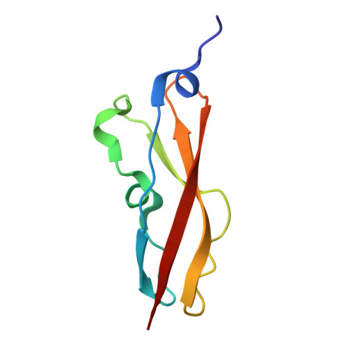
Crystal structure of the Rib domain of the cell-wall-anchored surface protein from Limosilactobacillus reuteri.

Xue, Y.Wu, Z.Kang, X.

(2024) Acta Crystallogr F Struct Biol Commun 80: 228-233

  • DOI: https://doi.org/10.1107/S2053230X24007970
  • Primary Citation of Related Structures:  
    8YK7

  • PubMed Abstract: 

    The immunoglobulin (Ig)-like domain is found in a broad range of proteins with diverse functional roles. While an essential β-sandwich fold is maintained, considerable structural variations exist and are critical for functional diversity. The Rib-domain family, primarily found as tandem-repeat modules in the surface proteins of Gram-positive bacteria, represents another significant structural variant of the Ig-like fold. However, limited structural and functional exploration of this family has been conducted, which significantly restricts the understanding of its evolution and significance within the Ig superclass. In this work, a high-resolution crystal structure of a Rib domain derived from the probiotic bacterium Limosilactobacillus reuteri is presented. This protein, while sharing significant structural similarity with homologous domains from other bacteria, exhibits a significantly increased thermal resistance. The potential structural features contributing to this stability are discussed. Moreover, the presence of two copper-binding sites, with one positioned on the interface, suggests potential functional roles that warrant further investigation.


  • Organizational Affiliation
    • Institute of Drug Discovery Technology, Ningbo University, Ningbo 315211, People's Republic of China.

Macromolecules
Find similar proteins by:  (by identity cutoff)  |  3D Structure
Entity ID: 1
MoleculeChains Sequence LengthOrganismDetailsImage
YSIRK signal domain/LPXTG anchor domain surface protein80Limosilactobacillus reuteriMutation(s): 0 
Gene Names: DB325_02015
UniProt
Find proteins for A0A2T5Q5G6 (Limosilactobacillus reuteri)
Explore A0A2T5Q5G6 
Go to UniProtKB:  A0A2T5Q5G6
Entity Groups  
Sequence Clusters30% Identity50% Identity70% Identity90% Identity95% Identity100% Identity
UniProt GroupA0A2T5Q5G6
Sequence Annotations
Expand
  • Reference Sequence
Experimental Data & Validation

Experimental Data

  • Method: X-RAY DIFFRACTION
  • Resolution: 1.35 Å
  • R-Value Free:  0.167 (Depositor), 0.173 (DCC) 
  • R-Value Work:  0.127 (Depositor), 0.133 (DCC) 
  • R-Value Observed: 0.129 (Depositor) 
Space Group: P 61
Unit Cell:
Length ( Å )Angle ( ˚ )
a = 76.274α = 90
b = 76.274β = 90
c = 23.326γ = 120
Software Package:
Software NamePurpose
REFMACrefinement
SAINTdata scaling
SADABSdata reduction
PHASERphasing

Structure Validation

View Full Validation Report


Entry History & Funding Information

Deposition Data


Funding OrganizationLocationGrant Number
National Natural Science Foundation of China (NSFC)China22074070

Revision History  (Full details and data files)

  • Version 1.0: 2024-09-11
    Type: Initial release
  • Version 1.1: 2024-09-18
    Changes: Database references