8YK3 | pdb_00008yk3

Blood group B alpha-1,3-galactosidase AgaBb from Bifidobacterium bifidum, construct T7-tag_24-673


Experimental Data Snapshot

  • Method: X-RAY DIFFRACTION
  • Resolution: 3.50 Å
  • R-Value Free: 
    0.285 (Depositor), 0.285 (DCC) 
  • R-Value Work: 
    0.203 (Depositor), 0.203 (DCC) 
  • R-Value Observed: 
    0.207 (Depositor) 

Starting Model: experimental
View more details

wwPDB Validation 3D Report Full Report

Validation slider image for 8YK3

Ligand Structure Quality Assessment 


This is version 1.1 of the entry. See complete history

Literature

Crystal Structure of Bifidobacterium bifidum Glycoside Hydrolase Family 110 alpha-Galactosidase Specific for Blood Group B Antigen.

Kashima, T.Akama, M.Wakinaka, T.Arakawa, T.Ashida, H.Fushinobu, S.

(2024) J Appl Glycosci (1999) 71: 81-90

  • DOI: https://doi.org/10.5458/jag.jag.JAG-2024_0005
  • Primary Citation Related Structures: 
    8YK1, 8YK2, 8YK3

  • PubMed Abstract: 

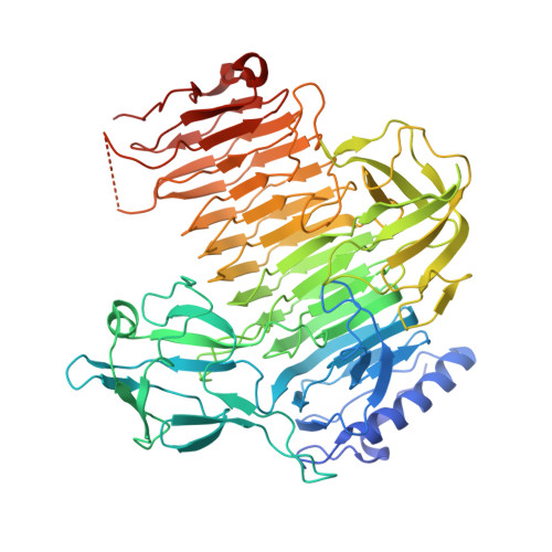
    To overcome incompatibility issues and increase the possibility of blood transfusion, technologies that enable efficient conversion of A- and B-type red blood cells to the universal donor O-type is desirable. Although several blood type-converting enzymes have been identified, detailed understanding about their molecular functions is limited. α-Galactosidase from Bifidobacterium bifidum JCM 1254 (AgaBb), belonging to glycoside hydrolase (GH) 110 subfamily A, specifically acts on blood group B antigen. Here we present the crystal structure of AgaBb, including the catalytic GH110 domain and part of the C-terminal uncharacterized regions. Based on this structure, we deduced a possible binding mechanism of blood group B antigen to the active site. Site-directed mutagenesis confirmed that R270 and E380 recognize the fucose moiety in the B antigen. Thermal shift assay revealed that the C-terminal uncharacterized region significantly contributes to protein stability. This region is shared only among GH110 enzymes from B. bifidum and some Ruminococcus species. The elucidation of the molecular basis for the specific recognition of blood group B antigen is expected to lead to the practical application of blood group conversion enzymes in the future.


  • Organizational Affiliation
    • 1 Department of Biotechnology, The University of Tokyo.

Macromolecule Content 

  • Total Structure Weight: 145.92 kDa 
  • Atom Count: 9,480 
  • Modeled Residue Count: 1,239 
  • Deposited Residue Count: 1,346 
  • Unique protein chains: 1

Macromolecules

Find similar proteins by:|  3D Structure
Entity ID: 1
MoleculeChains  Sequence LengthOrganismDetailsImage
Alpha-galactosidase
A, B
673Bifidobacterium bifidum JCM 1254Mutation(s): 0 
Gene Names: agabbGBA83_07165
EC: 3.2.1
UniProt
Find proteins for L8B3G2 (Bifidobacterium bifidum)
Explore L8B3G2 
Go to UniProtKB:  L8B3G2
Entity Groups
Sequence Clusters30% Identity50% Identity70% Identity90% Identity95% Identity100% Identity
UniProt GroupL8B3G2
Sequence Annotations
Expand
Reference Sequence

Small Molecules

Experimental Data & Validation

Experimental Data

  • Method: X-RAY DIFFRACTION
  • Resolution: 3.50 Å
  • R-Value Free:  0.285 (Depositor), 0.285 (DCC) 
  • R-Value Work:  0.203 (Depositor), 0.203 (DCC) 
  • R-Value Observed: 0.207 (Depositor) 
Space Group: P 1 21 1
Unit Cell:
Length ( Å )Angle ( ˚ )
a = 63.34α = 90
b = 73.154β = 91.38
c = 177.968γ = 90
Software Package:
Software NamePurpose
PHENIXrefinement
XDSdata reduction
Aimlessdata scaling
PHASERphasing

Structure Validation

View Full Validation Report


Ligand Structure Quality Assessment 


Entry History 

& Funding Information

Deposition Data


Funding OrganizationLocationGrant Number
Japan Society for the Promotion of Science (JSPS)Japan24380053
Japan Society for the Promotion of Science (JSPS)Japan23H00322

Revision History  (Full details and data files)

  • Version 1.0: 2024-07-10
    Type: Initial release
  • Version 1.1: 2024-09-18
    Changes: Database references