8YJR | pdb_00008yjr

Structure of the human endogenous PCNA-FEN1 complex - State D


Experimental Data Snapshot

  • Method: ELECTRON MICROSCOPY
  • Resolution: 3.51 Å
  • Aggregation State: PARTICLE 
  • Reconstruction Method: SINGLE PARTICLE 

wwPDB Validation 3D Report Full Report

Validation slider image for 8YJR

This is version 1.1 of the entry. See complete history

Literature

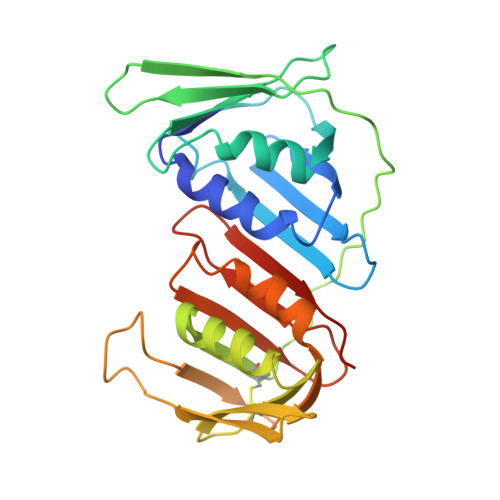
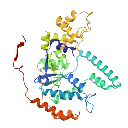
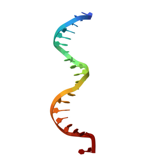
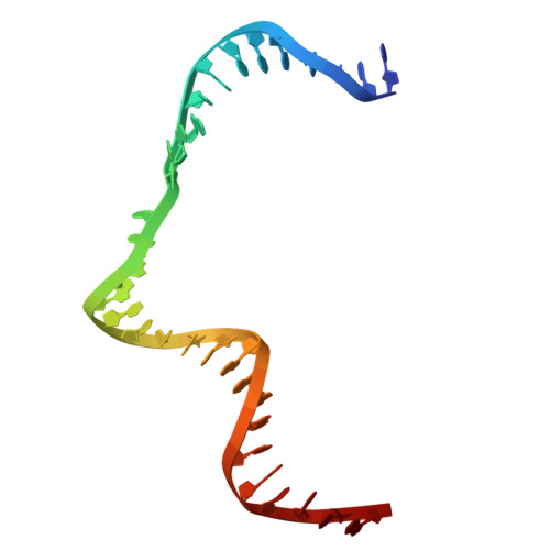
Structural insight into Okazaki fragment maturation mediated by PCNA-bound FEN1 and RNaseH2.

Tian, Y.Li, N.Li, Q.Gao, N.

(2025) EMBO J 44: 484-504

  • DOI: https://doi.org/10.1038/s44318-024-00296-x
  • Primary Citation Related Structures: 
    8YJH, 8YJL, 8YJQ, 8YJR, 8YJS, 8YJU, 8YJV, 8YJW, 8YJZ

  • PubMed Abstract: 

    PCNA is a master coordinator of many DNA-metabolic events. During DNA replication, the maturation of Okazaki fragments involves at least four DNA enzymes, all of which contain PCNA-interacting motifs. However, the temporal relationships and functional modulations between these PCNA-binding proteins are unclear. Here, we developed a strategy to purify endogenous PCNA-containing complexes from native chromatin, and characterized their structures using cryo-EM. Two structurally resolved classes (PCNA-FEN1 and PCNA-FEN1-RNaseH2 complexes) have captured a series of 3D snapshots for the primer-removal steps of Okazaki fragment maturation. These structures show that product release from FEN1 is a rate-liming step. Furthermore, both FEN1 and RNaseH2 undergo continuous conformational changes on PCNA that result in constant fluctuations in the bending angle of substrate DNA at the nick site, implying that these enzymes could regulate each other through conformational modulation of the bound DNA. The structures of the PCNA-FEN1-RNaseH2 complex confirm the toolbelt function of PCNA and suggests a potential unrecognized role of RNaseH2, as a dsDNA binding protein, in promoting the 5'-flap cleaving activity of FEN1.


  • Organizational Affiliation
    • State Key Laboratory of Membrane Biology, Peking-Tsinghua Joint Center for Life Sciences, School of Life Sciences, Peking University, Beijing, China.

Macromolecule Content 

  • Total Structure Weight: 148.92 kDa 
  • Atom Count: 9,962 
  • Modeled Residue Count: 1,178 
  • Deposited Residue Count: 1,228 
  • Unique protein chains: 2
  • Unique nucleic acid chains: 4

Macromolecules


Find similar proteins by:|  3D Structure
Entity ID: 1
MoleculeChains  Sequence LengthOrganismDetailsImage
Proliferating cell nuclear antigenA [auth B],
B [auth C],
D [auth A]
261Homo sapiensMutation(s): 0 
Gene Names: PCNA
UniProt & NIH Common Fund Data Resources
Find proteins for P12004 (Homo sapiens)
Explore P12004 
Go to UniProtKB:  P12004
PHAROS:  P12004
GTEx:  ENSG00000132646 
Entity Groups
Sequence Clusters30% Identity50% Identity70% Identity90% Identity95% Identity100% Identity
UniProt GroupP12004
Sequence Annotations
Expand
Reference Sequence
Find similar proteins by:|  3D Structure
Entity ID: 2
MoleculeChains  Sequence LengthOrganismDetailsImage
Flap endonuclease 1C [auth D]380Homo sapiensMutation(s): 0 
EC: 3.1
UniProt & NIH Common Fund Data Resources
Find proteins for P39748 (Homo sapiens)
Explore P39748 
Go to UniProtKB:  P39748
PHAROS:  P39748
GTEx:  ENSG00000168496 
Entity Groups
Sequence Clusters30% Identity50% Identity70% Identity90% Identity95% Identity100% Identity
UniProt GroupP39748
Sequence Annotations
Expand
Reference Sequence
Find similar nucleic acids by:  Sequence
Entity ID: 3
MoleculeChains LengthOrganismImage
upstream DNAE [auth J]20Homo sapiens
Sequence Annotations
Expand
Reference Sequence
Find similar nucleic acids by:  (by identity cutoff) 
Entity ID: 4
MoleculeChains LengthOrganismImage
parent DNAF [auth E]31Homo sapiens
Sequence Annotations
Expand
Reference Sequence
Find similar nucleic acids by:  Sequence
Entity ID: 5
MoleculeChains LengthOrganismImage
downstream DNAG [auth F]11Homo sapiens
Sequence Annotations
Expand
Reference Sequence
Find similar nucleic acids by:  Sequence
Entity ID: 6
MoleculeChains LengthOrganismImage
5 prime flap DNA3Homo sapiens
Sequence Annotations
Expand
Reference Sequence

Experimental Data & Validation

Experimental Data

  • Method: ELECTRON MICROSCOPY
  • Resolution: 3.51 Å
  • Aggregation State: PARTICLE 
  • Reconstruction Method: SINGLE PARTICLE 

Structure Validation

View Full Validation Report


Entry History 

& Funding Information

Deposition Data


Funding OrganizationLocationGrant Number
National Natural Science Foundation of China (NSFC)China--

Revision History  (Full details and data files)

  • Version 1.0: 2024-12-04
    Type: Initial release
  • Version 1.1: 2025-01-29
    Changes: Data collection, Database references