8YJ3 | pdb_00008yj3

N17.1.2 recognition of NRAS neoantigens


Experimental Data Snapshot

  • Method: X-RAY DIFFRACTION
  • Resolution: 3.50 Å
  • R-Value Free: 
    0.316 (Depositor), 0.311 (DCC) 
  • R-Value Work: 
    0.258 (Depositor), 0.257 (DCC) 
  • R-Value Observed: 
    0.261 (Depositor) 

Starting Model: experimental
View more details

wwPDB Validation 3D Report Full Report

Validation slider image for 8YJ3

This is version 1.0 of the entry. See complete history

Literature

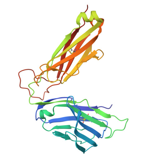
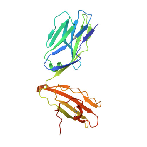
Structural characterization and AlphaFold modeling of human T cell receptor recognition of NRAS cancer neoantigens.

Wu, D.Yin, R.Chen, G.Ribeiro-Filho, H.V.Cheung, M.Robbins, P.F.Mariuzza, R.A.Pierce, B.G.

(2024) Sci Adv 10: eadq6150-eadq6150

  • DOI: https://doi.org/10.1126/sciadv.adq6150
  • Primary Citation Related Structures: 
    8YIV, 8YJ2, 8YJ3

  • PubMed Abstract: 

    T cell receptors (TCRs) that recognize cancer neoantigens are important for anticancer immune responses and immunotherapy. Understanding the structural basis of TCR recognition of neoantigens provides insights into their exquisite specificity and can enable design of optimized TCRs. We determined crystal structures of a human TCR in complex with NRAS Q61K and Q61R neoantigen peptides and HLA-A1 major histocompatibility complex (MHC), revealing the molecular underpinnings for dual recognition and specificity versus wild-type NRAS peptide. We then used multiple versions of AlphaFold to model the corresponding complex structures, given the challenge of immune recognition for such methods. One implementation of AlphaFold2 (TCRmodel2) with additional sampling was able to generate accurate models of the complexes, while AlphaFold3 also showed strong performance, although success was lower for other complexes. This study provides insights into TCR recognition of a shared cancer neoantigen as well as the utility and practical considerations for using AlphaFold to model TCR-peptide-MHC complexes.


  • Organizational Affiliation
    • Department of Hepatopancreatobiliary Surgery, The First Affiliated Hospital, Laboratory of Structural Immunology, Hengyang Medical School, University of South China, Hengyang, Hunan 421001, China.

Macromolecule Content 

  • Total Structure Weight: 205.05 kDa 
  • Atom Count: 14,248 
  • Modeled Residue Count: 1,796 
  • Deposited Residue Count: 1,816 
  • Unique protein chains: 2

Macromolecules

Find similar proteins by:|  3D Structure
Entity ID: 1
MoleculeChains  Sequence LengthOrganismDetailsImage
tcr betaA [auth E],
C [auth A],
E [auth C],
G
247Homo sapiensMutation(s): 0 
UniProt
Find proteins for P0DTU4 (Homo sapiens)
Explore P0DTU4 
Go to UniProtKB:  P0DTU4
Entity Groups
Sequence Clusters30% Identity50% Identity70% Identity90% Identity95% Identity100% Identity
UniProt GroupP0DTU4
Sequence Annotations
Expand
Reference Sequence
Find similar proteins by:|  3D Structure
Entity ID: 2
MoleculeChains  Sequence LengthOrganismDetailsImage
tcr alphaB [auth D],
D [auth B],
F,
H
207Homo sapiensMutation(s): 0 
UniProt & NIH Common Fund Data Resources
Find proteins for P01848 (Homo sapiens)
Explore P01848 
Go to UniProtKB:  P01848
PHAROS:  P01848
Entity Groups
Sequence Clusters30% Identity50% Identity70% Identity90% Identity95% Identity100% Identity
UniProt GroupP01848
Sequence Annotations
Expand
Reference Sequence

Experimental Data & Validation

Experimental Data

  • Method: X-RAY DIFFRACTION
  • Resolution: 3.50 Å
  • R-Value Free:  0.316 (Depositor), 0.311 (DCC) 
  • R-Value Work:  0.258 (Depositor), 0.257 (DCC) 
  • R-Value Observed: 0.261 (Depositor) 
Space Group: P 1 21 1
Unit Cell:
Length ( Å )Angle ( ˚ )
a = 103.228α = 90
b = 66.113β = 97.81
c = 138.59γ = 90
Software Package:
Software NamePurpose
PHENIXrefinement
HKL-2000data reduction
HKL-2000data scaling
PHENIXphasing

Structure Validation

View Full Validation Report


Entry History 

& Funding Information

Deposition Data


Funding OrganizationLocationGrant Number
National Natural Science Foundation of China (NSFC)China32270995
National Natural Science Foundation of China (NSFC)China32100985

Revision History  (Full details and data files)

  • Version 1.0: 2024-12-04
    Type: Initial release