


Structure of human PRPS2 long isoform at 3.4 Angstroms resolution.
Liu, J.L., Lu, G.M.To be published.
Experimental Data Snapshot
wwPDB Validation 3D Report Full Report

Entity ID: 1 | |||||
|---|---|---|---|---|---|
| Molecule | Chains | Sequence Length | Organism | Details | Image |
| Isoform 2 of Ribose-phosphate pyrophosphokinase 2 | 321 | Homo sapiens | Mutation(s): 0 Gene Names: PRPS2 EC: 2.7.6.1 | 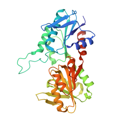 | |
UniProt & NIH Common Fund Data Resources | |||||
PHAROS: P11908 GTEx: ENSG00000101911 | |||||
Entity Groups | |||||
| Sequence Clusters | 30% Identity50% Identity70% Identity90% Identity95% Identity100% Identity | ||||
| UniProt Group | P11908 | ||||
Sequence AnnotationsExpand | |||||
Reference Sequence | |||||
| Ligands 3 Unique | |||||
|---|---|---|---|---|---|
| ID | Chains | Name / Formula / InChI Key | 2D Diagram | 3D Interactions | |
| ADP (Subject of Investigation/LOI) Download:Ideal Coordinates CCD File | AA [auth F] J [auth A] N [auth B] R [auth C] S [auth D] | ADENOSINE-5'-DIPHOSPHATE C10 H15 N5 O10 P2 XTWYTFMLZFPYCI-KQYNXXCUSA-N |  | ||
| RP5 (Subject of Investigation/LOI) Download:Ideal Coordinates CCD File | BA [auth F] G [auth A] K [auth B] O [auth C] T [auth D] | 5-O-phosphono-beta-D-ribofuranose C5 H11 O8 P KTVPXOYAKDPRHY-TXICZTDVSA-N |  | ||
| MG (Subject of Investigation/LOI) Download:Ideal Coordinates CCD File | CA [auth F] DA [auth F] H [auth A] I [auth A] L [auth B] | MAGNESIUM ION Mg JLVVSXFLKOJNIY-UHFFFAOYSA-N |  | ||
| Task | Software Package | Version |
|---|---|---|
| MODEL REFINEMENT | PHENIX | |
| Funding Organization | Location | Grant Number |
|---|---|---|
| National Natural Science Foundation of China (NSFC) | China | No. 31771490 |


