


The Crystal Structure of the Type I TGF beta receptor from Biortus.
Wang, F., Cheng, W., Lv, Z., Meng, Q., Xu, Y.To be published.
Experimental Data Snapshot
Starting Model: experimental
View more details
Entity ID: 1 | |||||
|---|---|---|---|---|---|
| Molecule | Chains | Sequence Length | Organism | Details | Image |
| TGF-beta receptor type-1 | 343 | Homo sapiens | Mutation(s): 0 Gene Names: TGFBR1, ALK5, SKR4 EC: 2.7.11.30 | 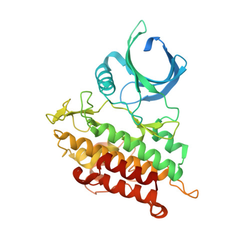 | |
UniProt & NIH Common Fund Data Resources | |||||
PHAROS: P36897 GTEx: ENSG00000106799 | |||||
Entity Groups | |||||
| Sequence Clusters | 30% Identity50% Identity70% Identity90% Identity95% Identity100% Identity | ||||
| UniProt Group | P36897 | ||||
Sequence AnnotationsExpand | |||||
Reference Sequence | |||||
| Ligands 1 Unique | |||||
|---|---|---|---|---|---|
| ID | Chains | Name / Formula / InChI Key | 2D Diagram | 3D Interactions | |
| A1D6I (Subject of Investigation/LOI) Download:Ideal Coordinates CCD File | B [auth A] | Vactosertib C22 H18 F N7 FJCDSQATIJKQKA-UHFFFAOYSA-N |  | ||
| Length ( Å ) | Angle ( ˚ ) |
|---|---|
| a = 42.196 | α = 90 |
| b = 76.764 | β = 90 |
| c = 89.707 | γ = 90 |
| Software Name | Purpose |
|---|---|
| REFMAC | refinement |
| XDS | data reduction |
| Aimless | data scaling |
| PHASER | phasing |
| Funding Organization | Location | Grant Number |
|---|---|---|
| Not funded | China | -- |


