


Crystal structure of L-ribulose 3-epimerase from Arthrobacter globiformis M30
Watanabe, M., Nakamich, Y., Mine, S.To be published.
Experimental Data Snapshot
Starting Model: experimental
View more details
Entity ID: 1 | |||||
|---|---|---|---|---|---|
| Molecule | Chains | Sequence Length | Organism | Details | Image |
| Ketose 3-epimerase | 297 | Arthrobacter globiformis | Mutation(s): 0 Gene Names: DAE EC: 5.1.3 | 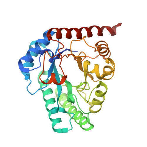 | |
UniProt | |||||
Find proteins for A0A1L7NQ96 (Arthrobacter globiformis) Explore A0A1L7NQ96 Go to UniProtKB: A0A1L7NQ96 | |||||
Entity Groups | |||||
| Sequence Clusters | 30% Identity50% Identity70% Identity90% Identity95% Identity100% Identity | ||||
| UniProt Group | A0A1L7NQ96 | ||||
Sequence AnnotationsExpand | |||||
Reference Sequence | |||||
| Ligands 2 Unique | |||||
|---|---|---|---|---|---|
| ID | Chains | Name / Formula / InChI Key | 2D Diagram | 3D Interactions | |
| PEG (Subject of Investigation/LOI) Download:Ideal Coordinates CCD File | AA [auth G], S [auth E], V [auth C], Y [auth F] | DI(HYDROXYETHYL)ETHER C4 H10 O3 MTHSVFCYNBDYFN-UHFFFAOYSA-N |  | ||
| MG (Subject of Investigation/LOI) Download:Ideal Coordinates CCD File | BA [auth H] CA [auth I] DA [auth J] EA [auth K] FA [auth L] | MAGNESIUM ION Mg JLVVSXFLKOJNIY-UHFFFAOYSA-N |  | ||
| Length ( Å ) | Angle ( ˚ ) |
|---|---|
| a = 130.095 | α = 90 |
| b = 148.264 | β = 94.97 |
| c = 136.707 | γ = 90 |
| Software Name | Purpose |
|---|---|
| REFMAC | refinement |
| XDS | data reduction |
| XDS | data scaling |
| PHASER | phasing |
| Funding Organization | Location | Grant Number |
|---|---|---|
| Not funded | -- |


