8YDC

Crystal structure of a hammerhead ribozyme with pseudoknot


Experimental Data Snapshot

  • Method: X-RAY DIFFRACTION
  • Resolution: 2.89 Å
  • R-Value Free: 0.209 
  • R-Value Work: 0.195 
  • R-Value Observed: 0.196 

wwPDB Validation   3D Report Full Report


Ligand Structure Quality Assessment 


This is version 1.0 of the entry. See complete history


Literature

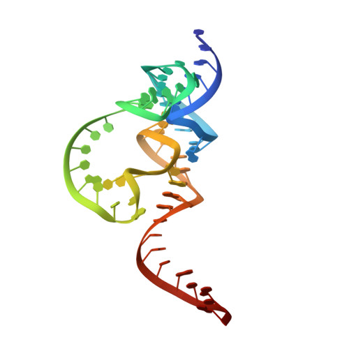
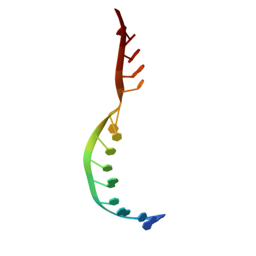
The structure and catalytic mechanism of a pseudoknot-containing hammerhead ribozyme.

Zhan, X.Wilson, T.J.Li, Z.Zhang, J.Yang, Y.Lilley, D.M.J.Liu, Y.

(2024) Nat Commun 15: 6628-6628

  • DOI: https://doi.org/10.1038/s41467-024-50892-y
  • Primary Citation of Related Structures:  
    8YDC

  • PubMed Abstract: 

    We have determined the crystal structure of a pseudoknot (PK)-containing hammerhead ribozyme that closely resembles the pistol ribozyme, with essentially identical secondary structure and connectivity. The activity is more sensitive to deletion of the G8 2'OH than to the absence of magnesium ions, indicating that the catalytic mechanism is the same as the extended hammerhead, and distinct from the pistol ribozyme. Here we show that nucleophilic attack is almost perfectly in-line, and the G8 2'OH is well positioned to act as general acid, being directed towards the O5' leaving group, and 2.9 Å away from it. Despite the similarity in overall structure to the pistol ribozyme, the local structure close to the cleavage site differs, and the PK hammerhead retains its unique mechanistic identity and demonstrates enhanced activity over other hammerhead ribozymes under standard conditions.


  • Organizational Affiliation

    State Key Laboratory of Medicinal Chemical Biology, Frontiers Science Center for Cell Responses, Nankai University, Tianjin, China.


Macromolecules
Find similar nucleic acids by: 3D Structure
Entity ID: 1
MoleculeChains LengthOrganismImage
ribozyme strand58synthetic construct
Sequence Annotations
Expand
  • Reference Sequence

Find similar nucleic acids by:  Sequence   |   3D Structure  

Entity ID: 2
MoleculeChains LengthOrganismImage
DNA/RNA (5'-R(*AP*CP*AP*UP*GP*UP*CP*U)-D(P*C)-R(P*UP*GP*GP*GP*A)-3')14synthetic construct
Sequence Annotations
Expand
  • Reference Sequence
Small Molecules
Ligands 1 Unique
IDChains Name / Formula / InChI Key2D Diagram3D Interactions
GTP (Subject of Investigation/LOI)
Query on GTP

Download Ideal Coordinates CCD File 
C [auth A]GUANOSINE-5'-TRIPHOSPHATE
C10 H16 N5 O14 P3
XKMLYUALXHKNFT-UUOKFMHZSA-N
Experimental Data & Validation

Experimental Data

  • Method: X-RAY DIFFRACTION
  • Resolution: 2.89 Å
  • R-Value Free: 0.209 
  • R-Value Work: 0.195 
  • R-Value Observed: 0.196 
  • Space Group: P 61 2 2
Unit Cell:
Length ( Å )Angle ( ˚ )
a = 83.578α = 90
b = 83.578β = 90
c = 258.79γ = 120
Software Package:
Software NamePurpose
PHENIXrefinement
DIALSdata reduction
DIALSdata scaling
PHENIXphasing

Structure Validation

View Full Validation Report


Ligand Structure Quality Assessment 


Entry History & Funding Information

Deposition Data


Funding OrganizationLocationGrant Number
National Science Foundation (NSF, China)China82111530210
National Science Foundation (NSF, China)China21877065

Revision History  (Full details and data files)

  • Version 1.0: 2024-08-21
    Type: Initial release