8Y33 | pdb_00008y33

A near-infrared fluorescent protein of de novo backbone design


Experimental Data Snapshot

  • Method: X-RAY DIFFRACTION
  • Resolution: 2.90 Å
  • R-Value Free: 
    0.244 (Depositor), 0.248 (DCC) 
  • R-Value Work: 
    0.200 (Depositor), 0.205 (DCC) 
  • R-Value Observed: 
    0.203 (Depositor) 

wwPDB Validation 3D Report Full Report

Validation slider image for 8Y33

Ligand Structure Quality Assessment 


This is version 1.2 of the entry. See complete history

Literature

Using Protein Design and Directed Evolution to Monomerize a Bright Near-Infrared Fluorescent Protein.

Hu, X.Xu, Y.Yi, J.Wang, C.Zhu, Z.Yue, T.Zhang, H.Wang, X.Wu, F.Xue, L.Bai, L.Liu, H.Chen, Q.

(2024) ACS Synth Biol 13: 1177-1190

  • DOI: https://doi.org/10.1021/acssynbio.3c00643
  • Primary Citation Related Structures: 
    8J1W, 8J1X, 8Y33

  • PubMed Abstract: 

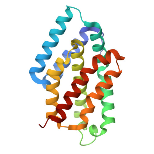
    The small ultrared fluorescent protein (smURFP) is a bright near-infrared (NIR) fluorescent protein (FP) that forms a dimer and binds its fluorescence chromophore, biliverdin, at its dimer interface. To engineer a monomeric NIR FP based on smURFP potentially more suitable for bioimaging, we employed protein design to extend the protein backbone with a new segment of two helices that shield the original dimer interface while covering the biliverdin binding pocket in place of the second chain in the original dimer. We experimentally characterized 13 designs and obtained a monomeric protein with a weak fluorescence. We enhanced the fluorescence of this designed protein through two rounds of directed evolution and obtained designed monomeric smURFP (DMsmURFP), a bright, stable, and monomeric NIR FP with a molecular weight of 19.6 kDa. We determined the crystal structures of DMsmURFP both in the apo state and in complex with biliverdin, which confirmed the designed structure. The use of DMsmURFP in in vivo imaging of mammalian systems was demonstrated. The backbone design-based strategy used here can also be applied to monomerize other naturally multimeric proteins with intersubunit functional sites.


  • Organizational Affiliation
    • Department of Rheumatology and Immunology, The First Affiliated Hospital of USTC, Center for Advanced Interdisciplinary Science and Biomedicine of IHM, Hefei National Center for Interdisciplinary Sciences at the Microscale, Division of Life Sciences and Medicine, University of Science and Technology of China, Hefei, Anhui 230027, China.

Macromolecule Content 

  • Total Structure Weight: 21.72 kDa 
  • Atom Count: 1,516 
  • Modeled Residue Count: 184 
  • Deposited Residue Count: 184 
  • Unique protein chains: 1

Macromolecules

Find similar proteins by:|  3D Structure
Entity ID: 1
MoleculeChains  Sequence LengthOrganismDetailsImage
near-infrared fluorescent protein184synthetic constructMutation(s): 0 

Small Molecules

Ligands 1 Unique
IDChains Name / Formula / InChI Key2D Diagram3D Interactions
V8U
(Subject of Investigation/LOI)

Query on V8U


Download:Ideal Coordinates CCD File
B [auth A]3-[5-[(4-ethenyl-3-methyl-5-oxidanylidene-pyrrol-2-yl)methyl]-2-[[5-[(3-ethyl-4-methyl-5-oxidanylidene-pyrrol-2-yl)methyl]-3-(3-hydroxy-3-oxopropyl)-4-methyl-1~{H}-pyrrol-2-yl]methyl]-4-methyl-1~{H}-pyrrol-3-yl]propanoic acid
C33 H38 N4 O6
UETWWFBMDBJSDH-UHFFFAOYSA-N

Experimental Data & Validation

Experimental Data

  • Method: X-RAY DIFFRACTION
  • Resolution: 2.90 Å
  • R-Value Free:  0.244 (Depositor), 0.248 (DCC) 
  • R-Value Work:  0.200 (Depositor), 0.205 (DCC) 
  • R-Value Observed: 0.203 (Depositor) 
Space Group: P 65
Unit Cell:
Length ( Å )Angle ( ˚ )
a = 105.987α = 90
b = 105.987β = 90
c = 41.691γ = 120
Software Package:
Software NamePurpose
PHENIXrefinement
XDSdata reduction
Aimlessdata scaling
MOLREPphasing

Structure Validation

View Full Validation Report


Ligand Structure Quality Assessment 


Entry History 

& Funding Information

Deposition Data

  • Released Date: 2024-02-28 
  • Deposition Author(s): Hu, X., Xu, Y.

Funding OrganizationLocationGrant Number
Not funded--

Revision History  (Full details and data files)

  • Version 1.0: 2024-02-28
    Type: Initial release
  • Version 1.1: 2024-04-17
    Changes: Database references
  • Version 1.2: 2024-05-01
    Changes: Database references