


Binding competition between substrates and cryoprotectants in protein crystals directly observed by a hybrid method of in-gel crystallization and high-pressure cryo-cooling in X-ray crystallography
Higashiura, A., Sugiyama, S.To be published.
Experimental Data Snapshot
Starting Model: experimental
View more details
Entity ID: 1 | |||||
|---|---|---|---|---|---|
| Molecule | Chains | Sequence Length | Organism | Details | Image |
| Streptavidin | 121 | Streptomyces avidinii | Mutation(s): 0 | 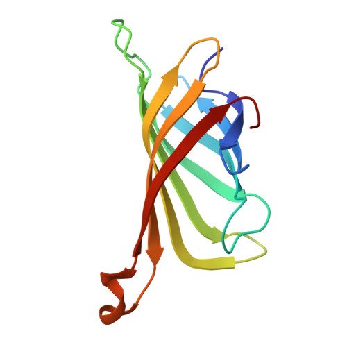 | |
UniProt | |||||
Entity Groups | |||||
| Sequence Clusters | 30% Identity50% Identity70% Identity90% Identity95% Identity100% Identity | ||||
| UniProt Group | P22629 | ||||
Sequence AnnotationsExpand | |||||
Reference Sequence | |||||
| Ligands 3 Unique | |||||
|---|---|---|---|---|---|
| ID | Chains | Name / Formula / InChI Key | 2D Diagram | 3D Interactions | |
| A1LXQ (Subject of Investigation/LOI) Download:Ideal Coordinates CCD File | G [auth B], H [auth C], K [auth D] | dimethyl 5-(4-oxidanylidene-5~{H}-furo[3,2-c]pyridin-2-yl)benzene-1,3-dicarboxylate C17 H13 N O6 RYAJKWVOTXPFPC-UHFFFAOYSA-N |  | ||
| GOL Download:Ideal Coordinates CCD File | F [auth A], I [auth C], J [auth C] | GLYCEROL C3 H8 O3 PEDCQBHIVMGVHV-UHFFFAOYSA-N |  | ||
| DMS Download:Ideal Coordinates CCD File | E [auth A] | DIMETHYL SULFOXIDE C2 H6 O S IAZDPXIOMUYVGZ-UHFFFAOYSA-N |  | ||
| Length ( Å ) | Angle ( ˚ ) |
|---|---|
| a = 46.988 | α = 90 |
| b = 85.539 | β = 98.92 |
| c = 58.367 | γ = 90 |
| Software Name | Purpose |
|---|---|
| PHENIX | refinement |
| XDS | data reduction |
| Aimless | data scaling |
| MOLREP | phasing |
| Funding Organization | Location | Grant Number |
|---|---|---|
| Other private | Japan | -- |


