8Y11

Crystal structure of L-2-keto-3-deoxyfuconate 4-dehydrogenase bound to NAD(H) and sulfate ion


Experimental Data Snapshot

  • Method: X-RAY DIFFRACTION
  • Resolution: 1.77 Å
  • R-Value Free: 0.211 
  • R-Value Work: 0.182 
  • R-Value Observed: 0.183 

Starting Model: in silico
View more details

wwPDB Validation   3D Report Full Report


Ligand Structure Quality Assessment 


This is version 1.0 of the entry. See complete history


Literature

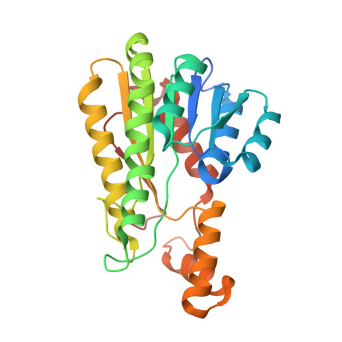
Crystal structure of L-2-keto-3-deoxyfuconate 4-dehydrogenase reveals a unique binding mode as a alpha-furanosyl hemiketal of substrates.

Akagashi, M.Watanabe, S.Kwiatkowski, S.Drozak, J.Terawaki, S.Watanabe, Y.

(2024) Sci Rep 14: 14602-14602

  • DOI: https://doi.org/10.1038/s41598-024-65627-8
  • Primary Citation of Related Structures:  
    8XWK, 8Y11, 8Y46, 8Y4B, 8Y4J

  • PubMed Abstract: 

    L-2-Keto-3-deoxyfuconate 4-dehydrogenase (L-KDFDH) catalyzes the NAD + -dependent oxidization of L-2-keto-3-deoxyfuconate (L-KDF) to L-2,4-diketo-3-deoxyfuconate (L-2,4-DKDF) in the non-phosphorylating L-fucose pathway from bacteria, and its substrate was previously considered to be the acyclic α-keto form of L-KDF. On the other hand, BDH2, a mammalian homolog with L-KDFDH, functions as a dehydrogenase for cis-4-hydroxy-L-proline (C4LHyp) with the cyclic structure. We found that L-KDFDH and BDH2 utilize C4LHyp and L-KDF, respectively. Therefore, to elucidate unique substrate specificity at the atomic level, we herein investigated for the first time the crystal structures of L-KDFDH from Herbaspirillum huttiense in the ligand-free, L-KDF and L-2,4-DKDF, D-KDP (D-2-keto-3-deoxypentonate; additional substrate), or L-2,4-DKDF and NADH bound forms. In complexed structures, L-KDF, L-2,4-DKDF, and D-KDP commonly bound as a α-furanosyl hemiketal. Furthermore, L-KDFDH showed no activity for L-KDF and D-KDP analogs without the C5 hydroxyl group, which form only the acyclic α-keto form. The C1 carboxyl and α-anomeric C2 hydroxyl groups and O5 oxygen atom of the substrate (and product) were specifically recognized by Arg148, Arg192, and Arg214. The side chain of Trp252 was important for hydrophobically recognizing the C6 methyl group of L-KDF. This is the first example showing the physiological role of the hemiketal of 2-keto-3-deoxysugar acid.


  • Organizational Affiliation

    Department of Bioscience, Graduate School of Agriculture, Ehime University, Matsuyama, Ehime, Japan.


Macromolecules
Find similar proteins by:  (by identity cutoff)  |  3D Structure
Entity ID: 1
MoleculeChains Sequence LengthOrganismDetailsImage
SDR family oxidoreductase
A, B, C, D
264Herbaspirillum huttienseMutation(s): 0 
Gene Names: E2K99_19890
Entity Groups  
Sequence Clusters30% Identity50% Identity70% Identity90% Identity95% Identity100% Identity
Sequence Annotations
Expand
  • Reference Sequence
Small Molecules
Ligands 5 Unique
IDChains Name / Formula / InChI Key2D Diagram3D Interactions
NAD (Subject of Investigation/LOI)
Query on NAD

Download Ideal Coordinates CCD File 
E [auth A],
I [auth B],
P [auth D]
NICOTINAMIDE-ADENINE-DINUCLEOTIDE
C21 H27 N7 O14 P2
BAWFJGJZGIEFAR-NNYOXOHSSA-N
TRS
Query on TRS

Download Ideal Coordinates CCD File 
M [auth B]2-AMINO-2-HYDROXYMETHYL-PROPANE-1,3-DIOL
C4 H12 N O3
LENZDBCJOHFCAS-UHFFFAOYSA-O
PEG
Query on PEG

Download Ideal Coordinates CCD File 
H [auth A],
L [auth B],
T [auth D],
U [auth D],
V [auth D]
DI(HYDROXYETHYL)ETHER
C4 H10 O3
MTHSVFCYNBDYFN-UHFFFAOYSA-N
SO4
Query on SO4

Download Ideal Coordinates CCD File 
F [auth A]
G [auth A]
J [auth B]
K [auth B]
N [auth C]
F [auth A],
G [auth A],
J [auth B],
K [auth B],
N [auth C],
Q [auth D],
R [auth D]
SULFATE ION
O4 S
QAOWNCQODCNURD-UHFFFAOYSA-L
GOL
Query on GOL

Download Ideal Coordinates CCD File 
O [auth C],
S [auth D]
GLYCEROL
C3 H8 O3
PEDCQBHIVMGVHV-UHFFFAOYSA-N
Experimental Data & Validation

Experimental Data

  • Method: X-RAY DIFFRACTION
  • Resolution: 1.77 Å
  • R-Value Free: 0.211 
  • R-Value Work: 0.182 
  • R-Value Observed: 0.183 
  • Space Group: P 21 21 21
Unit Cell:
Length ( Å )Angle ( ˚ )
a = 84.337α = 90
b = 103.894β = 90
c = 133.058γ = 90
Software Package:
Software NamePurpose
PHENIXrefinement
PDB_EXTRACTdata extraction
XDSdata scaling
PHASERphasing
XDSdata reduction

Structure Validation

View Full Validation Report


Ligand Structure Quality Assessment 


Entry History & Funding Information

Deposition Data


Funding OrganizationLocationGrant Number
Not funded--

Revision History  (Full details and data files)

  • Version 1.0: 2024-07-10
    Type: Initial release