Download Mendeley
Single-Molecule Insight Into alpha-Synuclein Fibril Structure and Mechanics Modulated by Chemical Compounds.
Li, X., Bi, L., Zhang, S., Xu, Q., Xia, W., Tao, Y., Wu, S., Li, Y., Le, W., Kang, W., Li, D., Sun, B., Liu, C.(2025) Adv Sci (Weinh) 12: e2416721-e2416721
- PubMed: 39951335 Search on PubMedSearch on PubMed Central
- DOI: https://doi.org/10.1002/advs.202416721
- Primary Citation Related Structures:
8XWD - PubMed Abstract:
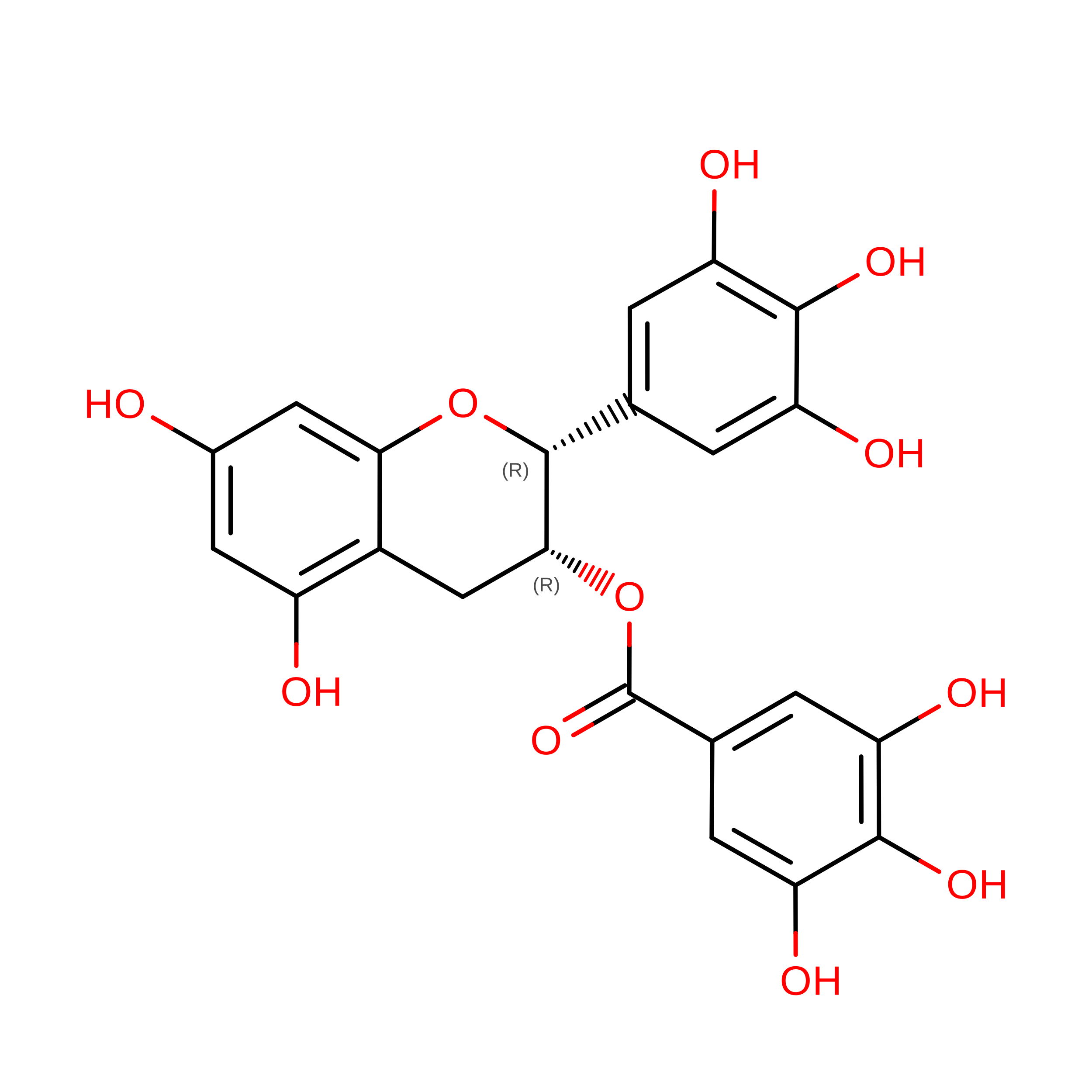
α-Syn fibrils, a key pathological hallmark of Parkinson's disease, is closely associated with disease initiation and progression. Several small molecules are found to bind or dissolve α-syn fibrils, offering potential therapeutic applications. Here, an innovative optical tweezers-based, fluorescence-combined approach is developed to probe the mechanical characteristics of α-syn fibrils at the single-molecule level. When subjected to axial stretching, local deformation within α-syn fibrils appeared at forces above 50 pN. These structural alternations occurred stepwise and are irreversible, suggesting unfolding of individual α-syn molecules or subdomains. Additionally, α-syn fibrils exhibits high heterogeneity in lateral disruption, with rupture force ranging from 50 to 500 pN. The impact of different compounds on the structure and mechanical features of α-syn fibrils is further examined. Notably, epigallocatechin gallate (EGCG) generally attenuates the rupture force of fibrils by wedging into the N-terminal polar groove and induces fibril dissociation. Conversely, copper chlorophyllin A (CCA) attaches to four different sites wrapping around the fibril core, reinforcing the stability of the fibril against rupture forces. The work offers an effective method for characterizing single-fibril properties and bridges compound-induced structural alternations with mechanical response. These insights are valuable for understanding amyloid fibril mechanics and their regulation by small molecules.
- Bio-X Institutes, Key Laboratory for the Genetics of Developmental and Neuropsychiatric Disorders (Ministry of Education), Shanghai Jiao Tong University, Shanghai, 200030, China.
Organizational Affiliation: