8XNJ | pdb_00008xnj

Crystal structure of trypsin in-complex with arginine


Experimental Data Snapshot

  • Method: X-RAY DIFFRACTION
  • Resolution: 2.40 Å
  • R-Value Free: 
    0.275 (Depositor), 0.269 (DCC) 
  • R-Value Work: 
    0.192 (Depositor), 0.199 (DCC) 
  • R-Value Observed: 
    0.196 (Depositor) 

Starting Model: experimental
View more details

wwPDB Validation 3D Report Full Report

Validation slider image for 8XNJ

Ligand Structure Quality Assessment 


This is version 1.0 of the entry. See complete history

Literature

In vitro , in silico and crystallographic-based identification of serine protease inhibitors.

Akbar, Z.Ahmad, M.S.

(2024) Nat Prod Res : 1-7

  • DOI: https://doi.org/10.1080/14786419.2024.2425793
  • Primary Citation Related Structures: 
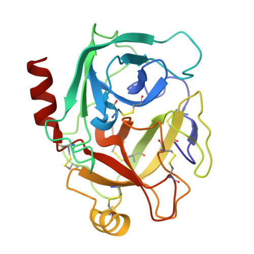
    8XNI, 8XNJ

  • PubMed Abstract: 

    Serine proteases are involved in various ailments, including pancreatitis, and colon cancer. Based on substrate recognition serine proteases are classified into different groups. Trypsin and trypsin-like serine proteases are among most studied group of serine proteases. Trypsin is among the chief hydrolysing enzyme involved in the pathogenesis of pancreatitis. Its inhibition can help to manage the disease. Herein, we investigated the trypsin inhibitory effect of some arginine-based small molecules, through in vitro , in silico , and crystallographic methods. Compounds 1 - 3 were evaluated against bovine pancreatic trypsin (BPT). Compound 1 was found to be active against trypsin with IC 50 value of 247.98 ± 2.44  μ M. Molecular docking studies were used to investigate the binding energy and binding conformation of inhibitor. All three compounds were subjected to crystallisation with trypsin. Compounds 1 - 2 were successfully crystallised with BPT The crystal structures of trypsin in complexed with compounds 1 , and 2 were determined at 2.30 and 2.50 Å resolution, respectively. Both molecules showed their binding affinity with the active site residues of trypsin. This study will provide insight into the binding mechanism of E-64 and arginine and might be useful in designing effective inhibitors of serine proteases.


  • Organizational Affiliation
    • H.E.J. Research Institute of Chemistry, International Center for Chemical and Biological Sciences, University of Karachi, Karachi, Pakistan.

Macromolecule Content 

  • Total Structure Weight: 23.75 kDa 
  • Atom Count: 1,750 
  • Modeled Residue Count: 223 
  • Deposited Residue Count: 223 
  • Unique protein chains: 1

Macromolecules

Find similar proteins by:|  3D Structure
Entity ID: 1
MoleculeChains  Sequence LengthOrganismDetailsImage
Cationic trypsin223Bos taurusMutation(s): 0 
EC: 3.4.21.4
UniProt
Find proteins for P00760 (Bos taurus)
Explore P00760 
Go to UniProtKB:  P00760
Entity Groups
Sequence Clusters30% Identity50% Identity70% Identity90% Identity95% Identity100% Identity
UniProt GroupP00760
Sequence Annotations
Expand
Reference Sequence

Small Molecules

Ligands 4 Unique
IDChains Name / Formula / InChI Key2D Diagram3D Interactions
ARG
(Subject of Investigation/LOI)

Query on ARG


Download:Ideal Coordinates CCD File
G [auth A]ARGININE
C6 H15 N4 O2
ODKSFYDXXFIFQN-BYPYZUCNSA-O
PEG

Query on PEG


Download:Ideal Coordinates CCD File
B [auth A]DI(HYDROXYETHYL)ETHER
C4 H10 O3
MTHSVFCYNBDYFN-UHFFFAOYSA-N
CA

Query on CA


Download:Ideal Coordinates CCD File
F [auth A]CALCIUM ION
Ca
BHPQYMZQTOCNFJ-UHFFFAOYSA-N
CL

Query on CL


Download:Ideal Coordinates CCD File
C [auth A],
D [auth A],
E [auth A]
CHLORIDE ION
Cl
VEXZGXHMUGYJMC-UHFFFAOYSA-M

Experimental Data & Validation

Experimental Data

  • Method: X-RAY DIFFRACTION
  • Resolution: 2.40 Å
  • R-Value Free:  0.275 (Depositor), 0.269 (DCC) 
  • R-Value Work:  0.192 (Depositor), 0.199 (DCC) 
  • R-Value Observed: 0.196 (Depositor) 
Space Group: P 21 21 21
Unit Cell:
Length ( Å )Angle ( ˚ )
a = 54.101α = 90
b = 58.152β = 90
c = 66.288γ = 90
Software Package:
Software NamePurpose
REFMACrefinement
PROTEUM PLUSdata reduction
Aimlessdata scaling
MOLREPphasing

Structure Validation

View Full Validation Report


Ligand Structure Quality Assessment 


Entry History 

& Funding Information

Deposition Data


Funding OrganizationLocationGrant Number
Not funded--

Revision History  (Full details and data files)

  • Version 1.0: 2024-12-04
    Type: Initial release