Funding Organization(s): Japan Agency for Medical Research and Development (AMED), Japan Science and Technology, Japan Society for the Promotion of Science (JSPS)
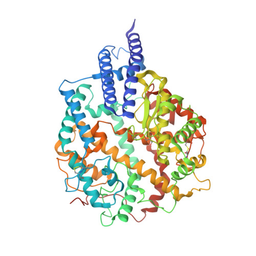
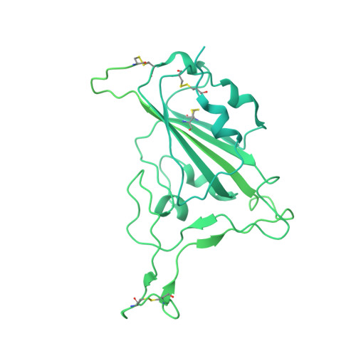
In middle to late 2023, a sublineage of severe acute respiratory syndrome coronavirus 2 (SARS-CoV-2) Omicron XBB, EG.5.1 (a progeny of XBB.1.9.2), is spreading rapidly around the world. We performed multiscale investigations, including phylogenetic analysis, epidemic dynamics modeling, infection experiments using pseudoviruses, clinical isolates, and recombinant viruses in cell cultures and experimental animals, and the use of human sera and antiviral compounds, to reveal the virological features of the newly emerging EG.5.1 variant. Our phylogenetic analysis and epidemic dynamics modeling suggested that two hallmark substitutions of EG.5.1, S:F456L and ORF9b:I5T are critical to its increased viral fitness. Experimental investigations on the growth kinetics, sensitivity to clinically available antivirals, fusogenicity, and pathogenicity of EG.5.1 suggested that the virological features of EG.5.1 are comparable to those of XBB.1.5. However, cryo-electron microscopy revealed structural differences between the spike proteins of EG.5.1 and XBB.1.5. We further assessed the impact of ORF9b:I5T on viral features, but it was almost negligible in our experimental setup. Our multiscale investigations provide knowledge for understanding the evolutionary traits of newly emerging pathogenic viruses, including EG.5.1, in the human population.
Organizational Affiliation:
Department of Microbiology and Immunology, Faculty of Medicine, Hokkaido University, Sapporo, Japan.
Center for iPS Cell Research and Application (CiRA), Kyoto University, Kyoto, Japan.
Laboratory of Biomolecular Science and Center for Research and Education on Drug Discovery, Faculty of Pharmaceutical Sciences, Hokkaido University, Sapporo, Japan.
First Medical Faculty at Biocev, Charles University, Vestec-Prague, Czechia.
Departamento de Farmacia, Facultad de Ciencias de la Salud, Universidad Cardenal Herrera-CEU (UCH-CEU), CEU Universities, Valencia, Spain.
Division of Systems Virology, Department of Microbiology and Immunology, The Institute of Medical Science, The University of Tokyo, Tokyo, Japan.
Department of Cancer Pathology, Faculty of Medicine, Hokkaido University, Sapporo, Japan.
Institute for Chemical Reaction Design and Discovery (WPI-ICReDD), Hokkaido University, Sapporo, Japan.
Division of Molecular Virology and Genetics, Joint Research Center for Human Retrovirus Infection, Kumamoto University, Kumamoto, Japan.
Graduate School of Medicine, The University of Tokyo, Tokyo, Japan.
Division of Risk Analysis and Management, International Institute for Zoonosis Control, Hokkaido University, Sapporo, Japan.
One Health Research Center, Hokkaido University, Sapporo, Japan.
Division of International Research Promotion, International Institute for Zoonosis Control, Hokkaido University, Sapporo, Japan.
Institute for Vaccine Research and Development (IVReD), Hokkaido University, Sapporo, Japan.
Department of Clinical Laboratory Medicine, Graduate School of Medicine, Kyoto University, Kyoto, Japan.
Laboratory of Medical Virology, Institute for Life and Medical Sciences, Kyoto University, Kyoto, Japan.
Department of Medicinal Sciences, Graduate School of Pharmaceutical Sciences, Kyushu University, Fukuoka, Japan.
Institute for Genetic Medicine, Hokkaido University, Sapporo, Japan.
HiLung Inc., Kyoto, Japan.
Tokyo Metropolitan Institute of Public Health, Tokyo, Japan.
Department of Clinical Pathology, Faculty of Medicine, Suez Canal University, Ismailia, Egypt.
Department of Biomedicine, School of Life Sciences, Indonesia International Institute for Life Sciences (i3L), Jakarta, Indonesia.
Department of Life Sciences, Faculty of Natural Science, Imperial College London, London, UK.
Graduate School of Frontier Sciences, The University of Tokyo, Kashiwa, Japan.
Global Station for Biosurfaces and Drug Discovery, Hokkaido University, Sapporo, Japan.
Division of Pathogen Structure, International Institute for Zoonosis Control, Hokkaido University, Sapporo, Japan.
Faculty of Pharmaceutical Sciences, Kyushu University, Fukuoka, Japan.
Graduate School of Biomedical and Health Sciences, Hiroshima University, Hiroshima, Japan.
International Collaboration Unit, International Institute for Zoonosis Control, Hokkaido University, Sapporo, Japan.
International Research Center for Infectious Diseases, The Institute of Medical Science, The University of Tokyo, Tokyo, Japan.
AMED-CREST, Japan Agency for Medical Research and Development (AMED), Tokyo, Japan.
CREST, Japan Science and Technology Agency, Kawaguchi, Japan.
Laboratory of Virus Control, Research Institute for Microbial Diseases, Osaka University, Suita, Japan.
International Vaccine Design Center, The Institute of Medical Science, The University of Tokyo, Tokyo, Japan.
Collaboration Unit for Infection, Joint Research Center for Human Retrovirus Infection, Kumamoto University, Kumamoto, Japan.