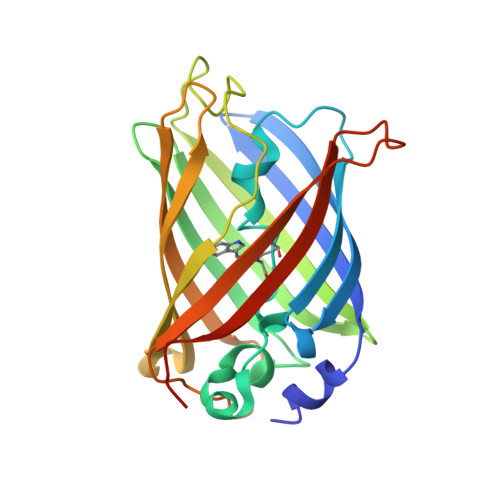
Comparison of two crystal polymorphs of NowGFP reveals a new conformational state trapped by crystal packing.
Kim, J.K., Jeong, H., Seo, J., Kim, S., Kim, K.H., Min, D., Kim, C.U.(2024) Acta Crystallogr D Struct Biol 80: 686-698
- PubMed: 39222305 Search on PubMed
- DOI: https://doi.org/10.1107/S2059798324008246
- Primary Citation Related Structures:
8XH0, 8XH1, 8XH2 - PubMed Abstract:
Crystal polymorphism serves as a strategy to study the conformational flexibility of proteins. However, the relationship between protein crystal packing and protein conformation often remains elusive. In this study, two distinct crystal forms of a green fluorescent protein variant, NowGFP, are compared: a previously identified monoclinic form (space group C2) and a newly discovered orthorhombic form (space group P2 1 2 1 2 1 ). Comparative analysis reveals that both crystal forms exhibit nearly identical linear assemblies of NowGFP molecules interconnected through similar crystal contacts. However, a notable difference lies in the stacking of these assemblies: parallel in the monoclinic form and perpendicular in the orthorhombic form. This distinct mode of stacking leads to different crystal contacts and induces structural alteration in one of the two molecules within the asymmetric unit of the orthorhombic crystal form. This new conformational state captured by orthorhombic crystal packing exhibits two unique features: a conformational shift of the β-barrel scaffold and a restriction of pH-dependent shifts of the key residue Lys61, which is crucial for the pH-dependent spectral shift of this protein. These findings demonstrate a clear connection between crystal packing and alternative conformational states of proteins, providing insights into how structural variations influence the function of fluorescent proteins.
- Department of Physics, Ulsan National Institute of Science and Technology (UNIST), Ulsan 44919, Republic of Korea.
Organizational Affiliation: