8XF2

Crystal structure of the reassembled C-phycocyanin hexamer from Thermoleptolyngbya sp. O-77


Experimental Data Snapshot

  • Method: X-RAY DIFFRACTION
  • Resolution: 1.98 Å
  • R-Value Free: 0.221 
  • R-Value Work: 0.183 
  • R-Value Observed: 0.183 

wwPDB Validation   3D Report Full Report


Ligand Structure Quality Assessment 


This is version 1.0 of the entry. See complete history


Literature

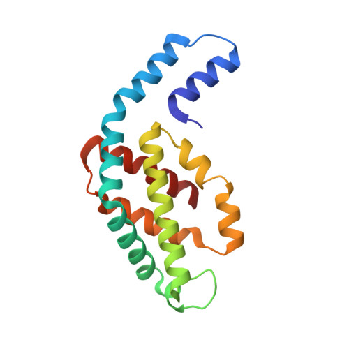
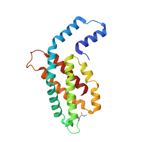
Disassembly and reassembly of the non-conventional thermophilic C-phycocyanin.

Nguyen, H.K.Minato, T.Teramoto, T.Ogo, S.Kakuta, Y.Yoon, K.S.

(2024) J Biosci Bioeng 137: 179-186

  • DOI: https://doi.org/10.1016/j.jbiosc.2023.12.015
  • Primary Citation of Related Structures:  
    8XF0, 8XF1, 8XF2, 8XF3

  • PubMed Abstract: 

    C-phycocyanin (CPC), which contains open-chain tetrapyrroles, is a major light-harvesting red-fluorescent protein with an important role in aquatic photosynthesis. Recently, we reported a non-conventional CPC from Thermoleptolyngbya sp. O-77 (CPC O77 ) that contains two different structures, i.e., a hexameric structure and a non-conventional octameric structure. However, the assembly and disassembly mechanisms of the non-conventional octameric form of CPC remain unclear. To understand this assembly mechanism, we performed an in vitro experiment to study the disassembly and reassembly behaviors of CPC using isolated CPC subunits. The dissociation of the CPC O77 subunit was performed using a Phenyl-Sepharose column in 20 mM potassium phosphate buffer (pH 6.0) containing 7.0 M urea. For the first time, crystals of isolated CPC subunits were obtained and analyzed after separation. After the removal of urea from the purified α and β subunits, we performed an in vitro reassembly experiment for CPC and analyzed the reconstructed CPC using spectrophotometric and X-ray crystal structure analyses. The crystal structure of the reassembled CPC was nearly identical to that of the original CPC O77 . The findings of this study indicate that the octameric CPC O77 is a naturally occurring form in the thermophilic cyanobacterium Thermoleptolyngbya sp. O-77.


  • Organizational Affiliation

    Department of Chemistry and Biochemistry, Graduate School of Engineering, Kyushu University, 744 Moto-oka, Nishi-ku, Fukuoka 819-0395, Japan.


Macromolecules
Find similar proteins by:  (by identity cutoff)  |  3D Structure
Entity ID: 1
MoleculeChains Sequence LengthOrganismDetailsImage
C-phycocyanin alpha chain
A, C, E, G, I
A, C, E, G, I, K
162Leptolyngbya sp. O-77Mutation(s): 0 
Entity Groups  
Sequence Clusters30% Identity50% Identity70% Identity90% Identity95% Identity100% Identity
Sequence Annotations
Expand
  • Reference Sequence
Find similar proteins by:  (by identity cutoff)  |  3D Structure
Entity ID: 2
MoleculeChains Sequence LengthOrganismDetailsImage
C-phycocyanin beta chain
B, D, F, H, J
B, D, F, H, J, L
172Leptolyngbya sp. O-77Mutation(s): 0 
Entity Groups  
Sequence Clusters30% Identity50% Identity70% Identity90% Identity95% Identity100% Identity
Sequence Annotations
Expand
  • Reference Sequence
Small Molecules
Ligands 3 Unique
IDChains Name / Formula / InChI Key2D Diagram3D Interactions
CYC
Query on CYC

Download Ideal Coordinates CCD File 
AA [auth G]
BA [auth H]
CA [auth H]
DA [auth I]
EA [auth J]
AA [auth G],
BA [auth H],
CA [auth H],
DA [auth I],
EA [auth J],
FA [auth J],
HA [auth K],
IA [auth L],
JA [auth L],
M [auth A],
O [auth B],
P [auth B],
Q [auth C],
S [auth D],
T [auth D],
V [auth E],
Y [auth F],
Z [auth F]
PHYCOCYANOBILIN
C33 H40 N4 O6
VXTXPYZGDQPMHK-GMXXPEQVSA-N
PO4
Query on PO4

Download Ideal Coordinates CCD File 
GA [auth J],
R [auth C],
X [auth E]
PHOSPHATE ION
O4 P
NBIIXXVUZAFLBC-UHFFFAOYSA-K
NA
Query on NA

Download Ideal Coordinates CCD File 
N [auth A],
U [auth D],
W [auth E]
SODIUM ION
Na
FKNQFGJONOIPTF-UHFFFAOYSA-N
Modified Residues  1 Unique
IDChains TypeFormula2D DiagramParent
MEN
Query on MEN
B, D, F, H, J
B, D, F, H, J, L
L-PEPTIDE LINKINGC5 H10 N2 O3ASN
Experimental Data & Validation

Experimental Data

  • Method: X-RAY DIFFRACTION
  • Resolution: 1.98 Å
  • R-Value Free: 0.221 
  • R-Value Work: 0.183 
  • R-Value Observed: 0.183 
  • Space Group: P 2 21 21
Unit Cell:
Length ( Å )Angle ( ˚ )
a = 107.729α = 90
b = 115.069β = 90
c = 186.989γ = 90
Software Package:
Software NamePurpose
PHENIXrefinement
XDSdata reduction
XDSdata scaling
PHASERphasing

Structure Validation

View Full Validation Report


Ligand Structure Quality Assessment 


Entry History & Funding Information

Deposition Data


Funding OrganizationLocationGrant Number
Japan Society for the Promotion of Science (JSPS)JapanJP26000008
Japan Society for the Promotion of Science (JSPS)JapanJP15K05566

Revision History  (Full details and data files)

  • Version 1.0: 2024-05-22
    Type: Initial release