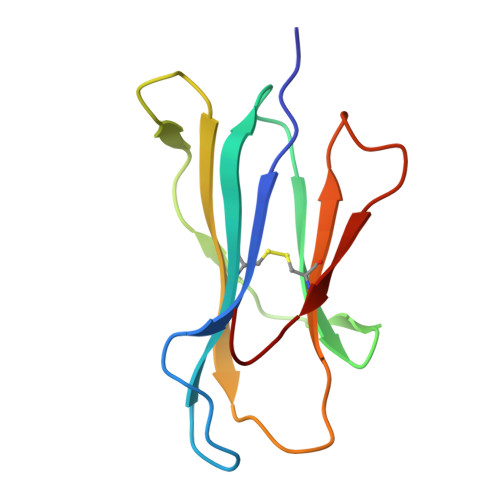
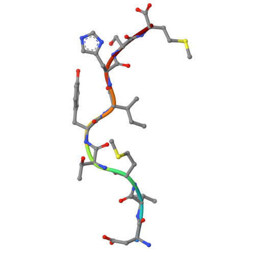
8XES

The structure of HLA-A/L1-1


Experimental Data Snapshot

  • Method: X-RAY DIFFRACTION
  • Resolution: 1.78 Å
  • R-Value Free: 0.235 
  • R-Value Work: 0.205 
  • R-Value Observed: 0.206 

Starting Model: experimental
View more details

wwPDB Validation   3D Report Full Report


This is version 1.1 of the entry. See complete history


Literature

Uncommon P1 Anchor-featured Viral T Cell Epitope Preference within HLA-A*2601 and HLA-A*0101 Individuals.

Zhang, J.Yue, C.Lin, Y.Tian, J.Guo, Y.Zhang, D.Guo, Y.Ye, B.Chai, Y.Qi, J.Zhao, Y.Gao, G.F.Sun, Z.Liu, J.

(2024) Immunohorizons 8: 415-430

  • DOI: https://doi.org/10.4049/immunohorizons.2400026
  • Primary Citation of Related Structures:  
    8XES, 8XFZ, 8XG2, 8XKC, 8XKE

  • PubMed Abstract: 

    The individual HLA-related susceptibility to emerging viral diseases such as COVID-19 underscores the importance of understanding how HLA polymorphism influences peptide presentation and T cell recognition. Similar to HLA-A*0101, which is one of the earliest identified HLA alleles among the human population, HLA-A*2601 possesses a similar characteristic for the binding peptide and acts as a prevalent allomorph in HLA-I. In this study, we found that, compared with HLA-A*0101, HLA-A*2601 individuals exhibit distinctive features for the T cell responses to SARS-CoV-2 and influenza virus after infection and/or vaccination. The heterogeneous T cell responses can be attributed to the distinct preference of HLA-A*2601 and HLA-A*0101 to T cell epitope motifs with negative-charged residues at the P1 and P3 positions, respectively. Furthermore, we determined the crystal structures of the HLA-A*2601 complexed to four peptides derived from SARS-CoV-2 and human papillomavirus, with one structure of HLA-A*0101 for comparison. The shallow pocket C of HLA-A*2601 results in the promiscuous presentation of peptides with "switchable" bulged conformations because of the secondary anchor in the median portion. Notably, the hydrogen bond network formed between the negative-charged P1 anchors and the HLA-A*2601-specific residues lead to a "closed" conformation and solid placement for the P1 secondary anchor accommodation in pocket A. This insight sheds light on the intricate relationship between HLA I allelic allomorphs, peptide binding, and the immune response and provides valuable implications for understanding disease susceptibility and potential vaccine design.


  • Organizational Affiliation

    National Key Laboratory of Intelligent Tracking and Forecasting for Infectious Diseases (NITFID), National Institute for Viral Disease Control and Prevention, Chinese Center for Disease Control and Prevention, Beijing, China.


Macromolecules
Find similar proteins by:  (by identity cutoff)  |  3D Structure
Entity ID: 1
MoleculeChains Sequence LengthOrganismDetailsImage
HLA class I heavy chain274Homo sapiensMutation(s): 0 
Gene Names: HLA-AHLAHLA-A26
UniProt
Find proteins for A0A0G2JPD3 (Homo sapiens)
Explore A0A0G2JPD3 
Go to UniProtKB:  A0A0G2JPD3
Entity Groups  
Sequence Clusters30% Identity50% Identity70% Identity90% Identity95% Identity100% Identity
UniProt GroupA0A0G2JPD3
Sequence Annotations
Expand
  • Reference Sequence
Find similar proteins by:  (by identity cutoff)  |  3D Structure
Entity ID: 2
MoleculeChains Sequence LengthOrganismDetailsImage
Beta-2-microglobulin99Homo sapiensMutation(s): 0 
Gene Names: B2MCDABP0092HDCMA22P
UniProt & NIH Common Fund Data Resources
Find proteins for P61769 (Homo sapiens)
Explore P61769 
Go to UniProtKB:  P61769
PHAROS:  P61769
GTEx:  ENSG00000166710 
Entity Groups  
Sequence Clusters30% Identity50% Identity70% Identity90% Identity95% Identity100% Identity
UniProt GroupP61769
Sequence Annotations
Expand
  • Reference Sequence

Find similar proteins by:  Sequence   |   3D Structure  

Entity ID: 3
MoleculeChains Sequence LengthOrganismDetailsImage
Major capsid protein L19Human papillomavirusMutation(s): 0 
UniProt
Find proteins for P03101 (Human papillomavirus type 16)
Explore P03101 
Go to UniProtKB:  P03101
Entity Groups  
UniProt GroupP03101
Sequence Annotations
Expand
  • Reference Sequence
Experimental Data & Validation

Experimental Data

  • Method: X-RAY DIFFRACTION
  • Resolution: 1.78 Å
  • R-Value Free: 0.235 
  • R-Value Work: 0.205 
  • R-Value Observed: 0.206 
  • Space Group: C 1 2 1
Unit Cell:
Length ( Å )Angle ( ˚ )
a = 167.116α = 90
b = 66.534β = 90.229
c = 49.327γ = 90
Software Package:
Software NamePurpose
PHENIXmodel building
PHENIXrefinement
PHASERphasing

Structure Validation

View Full Validation Report


Entry History & Funding Information

Deposition Data


Funding OrganizationLocationGrant Number
National Natural Science Foundation of China (NSFC)China--

Revision History  (Full details and data files)

  • Version 1.0: 2024-07-10
    Type: Initial release
  • Version 1.1: 2024-10-16
    Changes: Structure summary